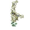
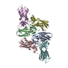
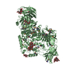
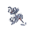
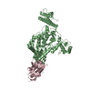
[English] 日本語
Yorodumi
- PDB-6oq8: Structure of the GTD domain of Clostridium difficile toxin B in c... -

+
Open data


ID or keywords:

Loading...

-
Basic information

Entry
Database: PDB / ID: 6oq8
TitleStructure of the GTD domain of Clostridium difficile toxin B in complex with VHH 7F
Components
  • 7F
  • Toxin B
KeywordsTOXIN / Toxin VHH
Function / homology
Function and homology information


glucosyltransferase activity / host cell cytosol / Transferases; Glycosyltransferases; Hexosyltransferases / cysteine-type peptidase activity / host cell endosome membrane / toxin activity / Hydrolases; Acting on peptide bonds (peptidases); Cysteine endopeptidases / lipid binding / host cell plasma membrane / proteolysis ...glucosyltransferase activity / host cell cytosol / Transferases; Glycosyltransferases; Hexosyltransferases / cysteine-type peptidase activity / host cell endosome membrane / toxin activity / Hydrolases; Acting on peptide bonds (peptidases); Cysteine endopeptidases / lipid binding / host cell plasma membrane / proteolysis / extracellular region / membrane / metal ion binding
Similarity search - Function
Arc Repressor Mutant, subunit A - #1780 / Methane Monooxygenase Hydroxylase; Chain G, domain 1 - #1190 / Dermonecrotic/RTX toxin, membrane localization domain / TcdA/TcdB toxin, N-terminal helical domain / TcdB toxin N-terminal helical domain / Membrane Localization Domain / TcdA/TcdB toxin, catalytic glycosyltransferase domain / TcdA/TcdB catalytic glycosyltransferase domain / TcdA/TcdB toxin, pore forming domain / TcdA/TcdB pore forming domain ...Arc Repressor Mutant, subunit A - #1780 / Methane Monooxygenase Hydroxylase; Chain G, domain 1 - #1190 / Dermonecrotic/RTX toxin, membrane localization domain / TcdA/TcdB toxin, N-terminal helical domain / TcdB toxin N-terminal helical domain / Membrane Localization Domain / TcdA/TcdB toxin, catalytic glycosyltransferase domain / TcdA/TcdB catalytic glycosyltransferase domain / TcdA/TcdB toxin, pore forming domain / TcdA/TcdB pore forming domain / CGT/MARTX, cysteine protease (CPD) domain / CGT/MARTX, cysteine protease (CPD) domain superfamily / Peptidase C80 family / CGT/MARTX cysteine protease (CPD) domain profile. / Choline-binding repeat / Putative cell wall binding repeat / Cell wall/choline-binding repeat / Cell wall-binding repeat profile. / Nucleotide-diphospho-sugar transferases / Methane Monooxygenase Hydroxylase; Chain G, domain 1 / Arc Repressor Mutant, subunit A / Up-down Bundle / Orthogonal Bundle / Mainly Alpha
Similarity search - Domain/homology
Biological speciesClostridioides difficile (bacteria)
Camelidae (mammal)
MethodX-RAY DIFFRACTION / SYNCHROTRON / MOLECULAR REPLACEMENT / Resolution: 2.2 Å
AuthorsChen, P. / Lam, K. / Jin, R.
Funding support United States, 1items
OrganizationGrant numberCountry
National Institutes of Health/National Center for Research Resources (NIH/NCRR) United States
CitationJournal: Nat.Struct.Mol.Biol. / Year: 2019
Title: Structure of the full-length Clostridium difficile toxin B.
Authors: Chen, P. / Lam, K.H. / Liu, Z. / Mindlin, F.A. / Chen, B. / Gutierrez, C.B. / Huang, L. / Zhang, Y. / Hamza, T. / Feng, H. / Matsui, T. / Bowen, M.E. / Perry, K. / Jin, R.
History
DepositionApr 25, 2019Deposition site: RCSB / Processing site: RCSB
Revision 1.0Jul 10, 2019Provider: repository / Type: Initial release
Revision 1.1Jul 31, 2019Group: Data collection / Database references / Category: citation / citation_author
Item: _citation.pdbx_database_id_PubMed / _citation.title / _citation_author.name
Revision 1.2Aug 21, 2019Group: Data collection / Database references / Category: citation
Item: _citation.journal_volume / _citation.page_first / _citation.page_last
Revision 1.3Dec 4, 2019Group: Author supporting evidence / Category: pdbx_audit_support / Item: _pdbx_audit_support.funding_organization
Revision 1.4Mar 13, 2024Group: Data collection / Database references / Refinement description
Category: chem_comp_atom / chem_comp_bond ...chem_comp_atom / chem_comp_bond / database_2 / struct_ncs_dom_lim
Item: _database_2.pdbx_DOI / _database_2.pdbx_database_accession ..._database_2.pdbx_DOI / _database_2.pdbx_database_accession / _struct_ncs_dom_lim.beg_auth_comp_id / _struct_ncs_dom_lim.beg_label_asym_id / _struct_ncs_dom_lim.beg_label_comp_id / _struct_ncs_dom_lim.beg_label_seq_id / _struct_ncs_dom_lim.end_auth_comp_id / _struct_ncs_dom_lim.end_label_asym_id / _struct_ncs_dom_lim.end_label_comp_id / _struct_ncs_dom_lim.end_label_seq_id

-
Structure visualization

Structure viewerMolecule:
MolmilJmol/JSmol

Downloads & links

-
Assembly

Deposited unit
A: Toxin B
D: 7F
B: Toxin B
C: 7F


Theoretical massNumber of molelcules
Total (without water)156,8594
Polymers156,8594
Non-polymers00
Water18,0331001
1
A: Toxin B
D: 7F


Theoretical massNumber of molelcules
Total (without water)78,4302
Polymers78,4302
Non-polymers00
Water362
TypeNameSymmetry operationNumber
identity operation1_555x,y,z1
Buried area1800 Å2
ΔGint-3 kcal/mol
Surface area30250 Å2
MethodPISA
2
B: Toxin B
C: 7F


Theoretical massNumber of molelcules
Total (without water)78,4302
Polymers78,4302
Non-polymers00
Water362
TypeNameSymmetry operationNumber
identity operation1_555x,y,z1
Buried area1800 Å2
ΔGint-5 kcal/mol
Surface area30440 Å2
MethodPISA
Unit cell
Length a, b, c (Å)114.711, 114.711, 301.010
Angle α, β, γ (deg.)90.00, 90.00, 90.00
Int Tables number96
Space group name H-MP43212
Noncrystallographic symmetry (NCS)NCS domain:
IDEns-IDDetails
11A
21B
12D
22C

NCS domain segments:

Component-ID: 0 / End auth comp-ID: SER / End label comp-ID: SER / Refine code: 0

Dom-IDEns-IDBeg auth comp-IDBeg label comp-IDAuth asym-IDLabel asym-IDAuth seq-IDLabel seq-ID
11GLYGLYAA0 - 5421 - 543
21GLYGLYBC0 - 5421 - 543
12SERSERDB-2 - 1151 - 118
22SERSERCD-2 - 1151 - 118

NCS ensembles :
ID
1
2

-
Components

#1: Protein Toxin B


Mass: 63129.512 Da / Num. of mol.: 2
Source method: isolated from a genetically manipulated source
Source: (gene. exp.) Clostridioides difficile (bacteria) / Gene: toxB, tcdB / Production host: Escherichia coli (E. coli)
References: UniProt: P18177, Hydrolases; Acting on peptide bonds (peptidases); Cysteine endopeptidases
#2: Antibody 7F


Mass: 15300.087 Da / Num. of mol.: 2
Source method: isolated from a genetically manipulated source
Source: (gene. exp.) Camelidae (mammal) / Production host: Escherichia coli (E. coli)
#3: Water ChemComp-HOH / water / Water


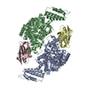
Mass: 18.015 Da / Num. of mol.: 1001 / Source method: isolated from a natural source / Formula: H2O

-
Experimental details

-
Experiment

ExperimentMethod: X-RAY DIFFRACTION / Number of used crystals: 1

-
Sample preparation

CrystalDensity Matthews: 3.16 Å3/Da / Density % sol: 61.03 %
Crystal growTemperature: 291 K / Method: vapor diffusion, hanging drop
Details: 0.1M Tris, pH 8.8, 0.4 M ammonium sulfate, and 24% PEG 3350

-
Data collection

DiffractionMean temperature: 100 K / Serial crystal experiment: N
Diffraction sourceSource: SYNCHROTRON / Site: APS / Beamline: 24-ID-C / Wavelength: 0.97919 Å
DetectorType: DECTRIS PILATUS 6M-F / Detector: PIXEL / Date: Apr 11, 2017
RadiationProtocol: SINGLE WAVELENGTH / Monochromatic (M) / Laue (L): M / Scattering type: x-ray
Radiation wavelengthWavelength: 0.97919 Å / Relative weight: 1
ReflectionResolution: 2.2→107.19 Å / Num. obs: 112567 / % possible obs: 99.6 % / Redundancy: 13.4 % / Rmerge(I) obs: 0.11 / Net I/σ(I): 19.2
Reflection shellResolution: 2.2→2.26 Å / Rmerge(I) obs: 0.63 / Num. unique obs: 9996

-
Processing

Software
NameVersionClassification
REFMAC5.8.0232refinement
XDSdata reduction
XDSdata scaling
PHASERphasing
RefinementMethod to determine structure: MOLECULAR REPLACEMENT / Resolution: 2.2→107.19 Å / Cor.coef. Fo:Fc: 0.947 / Cor.coef. Fo:Fc free: 0.935 / SU B: 4.758 / SU ML: 0.121 / Cross valid method: THROUGHOUT / ESU R: 0.214 / ESU R Free: 0.171
RfactorNum. reflection% reflectionSelection details
Rfree0.21989 5157 5 %RANDOM
Rwork0.19736 ---
obs0.19853 97145 99.63 %-
Solvent computationIon probe radii: 0.8 Å / Shrinkage radii: 0.8 Å / VDW probe radii: 1.2 Å
Displacement parametersBiso mean: 32.767 Å2
Baniso -1Baniso -2Baniso -3
1-0.1 Å20 Å20 Å2
2--0.1 Å20 Å2
3----0.19 Å2
Refinement stepCycle: 1 / Resolution: 2.2→107.19 Å
ProteinNucleic acidLigandSolventTotal
Num. atoms10683 0 0 1001 11684
Refine LS restraints
Refine-IDTypeDev idealDev ideal targetNumber
X-RAY DIFFRACTIONr_bond_refined_d0.0030.01210965
X-RAY DIFFRACTIONr_bond_other_d
X-RAY DIFFRACTIONr_angle_refined_deg0.8981.63314808
X-RAY DIFFRACTIONr_angle_other_deg
X-RAY DIFFRACTIONr_dihedral_angle_1_deg5.66851333
X-RAY DIFFRACTIONr_dihedral_angle_2_deg33.46924.146615
X-RAY DIFFRACTIONr_dihedral_angle_3_deg14.388152000
X-RAY DIFFRACTIONr_dihedral_angle_4_deg20.3471550
X-RAY DIFFRACTIONr_chiral_restr0.0820.21423
X-RAY DIFFRACTIONr_gen_planes_refined0.0030.028396
X-RAY DIFFRACTIONr_gen_planes_other
X-RAY DIFFRACTIONr_nbd_refined
X-RAY DIFFRACTIONr_nbd_other
X-RAY DIFFRACTIONr_nbtor_refined
X-RAY DIFFRACTIONr_nbtor_other
X-RAY DIFFRACTIONr_xyhbond_nbd_refined
X-RAY DIFFRACTIONr_xyhbond_nbd_other
X-RAY DIFFRACTIONr_metal_ion_refined
X-RAY DIFFRACTIONr_metal_ion_other
X-RAY DIFFRACTIONr_symmetry_vdw_refined
X-RAY DIFFRACTIONr_symmetry_vdw_other
X-RAY DIFFRACTIONr_symmetry_hbond_refined
X-RAY DIFFRACTIONr_symmetry_hbond_other
X-RAY DIFFRACTIONr_symmetry_metal_ion_refined
X-RAY DIFFRACTIONr_symmetry_metal_ion_other
X-RAY DIFFRACTIONr_mcbond_it1.2353.1335326
X-RAY DIFFRACTIONr_mcbond_other
X-RAY DIFFRACTIONr_mcangle_it2.1414.6936661
X-RAY DIFFRACTIONr_mcangle_other
X-RAY DIFFRACTIONr_scbond_it1.5533.285639
X-RAY DIFFRACTIONr_scbond_other
X-RAY DIFFRACTIONr_scangle_it
X-RAY DIFFRACTIONr_scangle_other
X-RAY DIFFRACTIONr_long_range_B_refined4.8641.97717150
X-RAY DIFFRACTIONr_long_range_B_other
X-RAY DIFFRACTIONr_rigid_bond_restr
X-RAY DIFFRACTIONr_sphericity_free
X-RAY DIFFRACTIONr_sphericity_bonded
Refine LS restraints NCS

Refine-ID: X-RAY DIFFRACTION / Type: interatomic distance / Weight position: 0.05

Ens-IDDom-IDAuth asym-IDNumberRms dev position (Å)
11A188730.06
12B188730.06
21D35070.1
22C35070.1
LS refinement shellResolution: 2.2→2.257 Å / Total num. of bins used: 20
RfactorNum. reflection% reflection
Rfree0.255 379 -
Rwork0.221 7028 -
obs--99.13 %

+
About Yorodumi

-
News

-
Feb 9, 2022. New format data for meta-information of EMDB entries

New format data for meta-information of EMDB entries

  • Version 3 of the EMDB header file is now the official format.
  • The previous official version 1.9 will be removed from the archive.

Related info.:EMDB header

External links:wwPDB to switch to version 3 of the EMDB data model

-
Aug 12, 2020. Covid-19 info

Covid-19 info

URL: https://pdbj.org/emnavi/covid19.php

New page: Covid-19 featured information page in EM Navigator.

Related info.:Covid-19 info / Mar 5, 2020. Novel coronavirus structure data

+
Mar 5, 2020. Novel coronavirus structure data

Novel coronavirus structure data

Related info.:Yorodumi Speices / Aug 12, 2020. Covid-19 info

External links:COVID-19 featured content - PDBj / Molecule of the Month (242):Coronavirus Proteases

+
Jan 31, 2019. EMDB accession codes are about to change! (news from PDBe EMDB page)

EMDB accession codes are about to change! (news from PDBe EMDB page)

  • The allocation of 4 digits for EMDB accession codes will soon come to an end. Whilst these codes will remain in use, new EMDB accession codes will include an additional digit and will expand incrementally as the available range of codes is exhausted. The current 4-digit format prefixed with “EMD-” (i.e. EMD-XXXX) will advance to a 5-digit format (i.e. EMD-XXXXX), and so on. It is currently estimated that the 4-digit codes will be depleted around Spring 2019, at which point the 5-digit format will come into force.
  • The EM Navigator/Yorodumi systems omit the EMD- prefix.

Related info.:Q: What is EMD? / ID/Accession-code notation in Yorodumi/EM Navigator

External links:EMDB Accession Codes are Changing Soon! / Contact to PDBj

+
Jul 12, 2017. Major update of PDB

Major update of PDB

  • wwPDB released updated PDB data conforming to the new PDBx/mmCIF dictionary.
  • This is a major update changing the version number from 4 to 5, and with Remediation, in which all the entries are updated.
  • In this update, many items about electron microscopy experimental information are reorganized (e.g. em_software).
  • Now, EM Navigator and Yorodumi are based on the updated data.

External links:wwPDB Remediation / Enriched Model Files Conforming to OneDep Data Standards Now Available in the PDB FTP Archive

-
Yorodumi

Thousand views of thousand structures

  • Yorodumi is a browser for structure data from EMDB, PDB, SASBDB, etc.
  • This page is also the successor to EM Navigator detail page, and also detail information page/front-end page for Omokage search.
  • The word "yorodu" (or yorozu) is an old Japanese word meaning "ten thousand". "mi" (miru) is to see.

Related info.:EMDB / PDB / SASBDB / Comparison of 3 databanks / Yorodumi Search / Aug 31, 2016. New EM Navigator & Yorodumi / Yorodumi Papers / Jmol/JSmol / Function and homology information / Changes in new EM Navigator and Yorodumi

Read more