 Movie
Movie Controller

[English] 日本語
 Yorodumi
- PDB-6g59: Structure of the alanine racemase from Staphylococcus aureus in c... -
+ Open data

- Basic information
| Entry | Database: PDB / ID: 6g59 | |||||||||
|---|---|---|---|---|---|---|---|---|---|---|
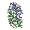
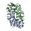
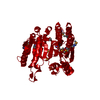
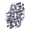
| Title | Structure of the alanine racemase from Staphylococcus aureus in complex with an pyridoxal-6- phosphate derivative | |||||||||
 Components Components | Alanine racemase 1 | |||||||||
 Keywords Keywords | BIOSYNTHETIC PROTEIN / pyridoxal 5 phosphate dependent / D-alanine biosynthesis | |||||||||
| Function / homology |  Function and homology information Function and homology informationalanine racemase / D-alanine biosynthetic process / alanine racemase activity / peptidoglycan biosynthetic process / pyridoxal phosphate binding / cytosol Similarity search - Function | |||||||||
| Biological species |  Staphylococcus aureus subsp. aureus Mu50 (bacteria) Staphylococcus aureus subsp. aureus Mu50 (bacteria) | |||||||||
| Method |  X-RAY DIFFRACTION / SYNCHROTRON / MOLECULAR REPLACEMENT / Resolution: 2.45 Å X-RAY DIFFRACTION / SYNCHROTRON / MOLECULAR REPLACEMENT / Resolution: 2.45 Å | |||||||||
 Authors Authors | Hoegl, A. / Sieber, S.A. / Schneider, S. | |||||||||
| Funding support |  Germany, 2items Germany, 2items
| |||||||||
 Citation Citation | Journal: Nat Chem / Year: 2018 Title: Mining the cellular inventory of pyridoxal phosphate-dependent enzymes with functionalized cofactor mimics. Authors: Hoegl, A. / Nodwell, M.B. / Kirsch, V.C. / Bach, N.C. / Pfanzelt, M. / Stahl, M. / Schneider, S. / Sieber, S.A. | |||||||||
| History |
|
- Structure visualization
| Structure viewer | Molecule: MolmilJmol/JSmol |
|---|
- Downloads & links
-Download
| PDBx/mmCIF format | 6g59.cif.gz | 317.1 KB | Display | PDBx/mmCIF format |
|---|---|---|---|---|
| PDB format | pdb6g59.ent.gz | 257 KB | Display | PDB format |
| PDBx/mmJSON format | 6g59.json.gz | Tree view | PDBx/mmJSON format | |
| Others |  Other downloads Other downloads |
-Validation report
| Arichive directory | https://data.pdbj.org/pub/pdb/validation_reports/g5/6g59ftp://data.pdbj.org/pub/pdb/validation_reports/g5/6g59 | HTTPS FTP |
|---|
-Related structure data
| Related structure data |  6g56C  6g58C  4a3qS S: Starting model for refinement C: citing same article ( |
|---|---|
| Similar structure data |


-Links
 PDBj
PDBj- Assembly
| Deposited unit | 
| ||||||||||||||||||
|---|---|---|---|---|---|---|---|---|---|---|---|---|---|---|---|---|---|---|---|
| 1 |
| ||||||||||||||||||
| Unit cell |
| ||||||||||||||||||
| Noncrystallographic symmetry (NCS) | NCS domain:
NCS domain segments: Component-ID: _ / Ens-ID: 1 / Beg auth comp-ID: ALA / Beg label comp-ID: ALA / End auth comp-ID: LEU / End label comp-ID: LEU / Refine code: _ / Auth seq-ID: -1 - 381 / Label seq-ID: 21 - 403
|
-Components
-Protein , 1 types, 2 molecules AB
| #1: Protein | Mass: 45285.801 Da / Num. of mol.: 2 Source method: isolated from a genetically manipulated source Details: N-terminal strep-tag Source: (gene. exp.) Staphylococcus aureus subsp. aureus Mu50 (bacteria)Gene: alr1, alr, SAV2070 / Production host: |
|---|
-Non-polymers , 5 types, 185 molecules 


| #2: Chemical |  Mass: 257.137 Da / Num. of mol.: 2 / Source method: obtained synthetically / Formula: C9H8NO6P Mass: 257.137 Da / Num. of mol.: 2 / Source method: obtained synthetically / Formula: C9H8NO6P#3: Chemical | ChemComp-SO4 /  Mass: 96.063 Da / Num. of mol.: 6 / Source method: obtained synthetically / Formula: SO4 Mass: 96.063 Da / Num. of mol.: 6 / Source method: obtained synthetically / Formula: SO4#4: Chemical | ChemComp-NA /  Mass: 22.990 Da / Num. of mol.: 4 / Source method: obtained synthetically / Formula: Na Mass: 22.990 Da / Num. of mol.: 4 / Source method: obtained synthetically / Formula: Na#5: Chemical |  Mass: 35.453 Da / Num. of mol.: 2 / Source method: obtained synthetically / Formula: Cl Mass: 35.453 Da / Num. of mol.: 2 / Source method: obtained synthetically / Formula: Cl#6: Water | ChemComp-HOH / | Mass: 18.015 Da / Num. of mol.: 171 / Source method: isolated from a natural source / Formula: H2O |
|---|
-Details
| Has protein modification | Y |
|---|
-Experimental details
-Experiment
| Experiment | Method: X-RAY DIFFRACTION / Number of used crystals: 1 |
|---|
- Sample preparation
| Crystal | Density Matthews: 3.32 Å3/Da / Density % sol: 62.96 % |
|---|---|
| Crystal grow | Temperature: 277 K / Method: vapor diffusion / Details: 0.1M Hepes pH7, 2M NaCl |
-Data collection
| Diffraction | Mean temperature: 100 K |
|---|---|
| Diffraction source | Source: SYNCHROTRON / Site: ESRF  / Beamline: ID23-2 / Wavelength: 0.873 Å / Beamline: ID23-2 / Wavelength: 0.873 Å |
| Detector | Type: DECTRIS PILATUS3 2M / Detector: PIXEL / Date: May 15, 2012 |
| Radiation | Protocol: SINGLE WAVELENGTH / Monochromatic (M) / Laue (L): M / Scattering type: x-ray |
| Radiation wavelength | Wavelength: 0.873 Å / Relative weight: 1 |
| Reflection | Resolution: 2.45→49.5 Å / Num. obs: 44955 / % possible obs: 100 % / Redundancy: 5 % / Biso Wilson estimate: 32.3 Å2 / CC1/2: 0.98 / Rmerge(I) obs: 0.2 / Net I/σ(I): 6.6 |
| Reflection shell | Resolution: 2.45→2.54 Å / Redundancy: 4.9 % / Rmerge(I) obs: 1 / Mean I/σ(I) obs: 1.4 / Num. unique obs: 4399 / CC1/2: 0.5 / % possible all: 99 |
- Processing
| Software |
| ||||||||||||||||||||||||||||||||||||||||||||||||||||||||||||||||||||||||||||||||||||||||||||||||||||||||||||||||||||||||||||||||||||||||||||||||||||||||||||||||||||||||||||||||||||||
|---|---|---|---|---|---|---|---|---|---|---|---|---|---|---|---|---|---|---|---|---|---|---|---|---|---|---|---|---|---|---|---|---|---|---|---|---|---|---|---|---|---|---|---|---|---|---|---|---|---|---|---|---|---|---|---|---|---|---|---|---|---|---|---|---|---|---|---|---|---|---|---|---|---|---|---|---|---|---|---|---|---|---|---|---|---|---|---|---|---|---|---|---|---|---|---|---|---|---|---|---|---|---|---|---|---|---|---|---|---|---|---|---|---|---|---|---|---|---|---|---|---|---|---|---|---|---|---|---|---|---|---|---|---|---|---|---|---|---|---|---|---|---|---|---|---|---|---|---|---|---|---|---|---|---|---|---|---|---|---|---|---|---|---|---|---|---|---|---|---|---|---|---|---|---|---|---|---|---|---|---|---|---|---|
| Refinement | Method to determine structure: MOLECULAR REPLACEMENT Starting model: 4A3Q Resolution: 2.45→49.5 Å / Cor.coef. Fo:Fc: 0.941 / Cor.coef. Fo:Fc free: 0.93 / SU B: 15.869 / SU ML: 0.171 / Cross valid method: THROUGHOUT / ESU R: 0.29 / ESU R Free: 0.206 / Details: HYDROGENS HAVE BEEN ADDED IN THE RIDING POSITIONS
| ||||||||||||||||||||||||||||||||||||||||||||||||||||||||||||||||||||||||||||||||||||||||||||||||||||||||||||||||||||||||||||||||||||||||||||||||||||||||||||||||||||||||||||||||||||||
| Solvent computation | Ion probe radii: 0.8 Å / Shrinkage radii: 0.8 Å / VDW probe radii: 1.1 Å | ||||||||||||||||||||||||||||||||||||||||||||||||||||||||||||||||||||||||||||||||||||||||||||||||||||||||||||||||||||||||||||||||||||||||||||||||||||||||||||||||||||||||||||||||||||||
| Displacement parameters | Biso mean: 35.447 Å2
| ||||||||||||||||||||||||||||||||||||||||||||||||||||||||||||||||||||||||||||||||||||||||||||||||||||||||||||||||||||||||||||||||||||||||||||||||||||||||||||||||||||||||||||||||||||||
| Refinement step | Cycle: 1 / Resolution: 2.45→49.5 Å
| ||||||||||||||||||||||||||||||||||||||||||||||||||||||||||||||||||||||||||||||||||||||||||||||||||||||||||||||||||||||||||||||||||||||||||||||||||||||||||||||||||||||||||||||||||||||
| Refine LS restraints |
|