Resolution: 2.3→2.34 Å / Redundancy: 7.8 % / Rmerge(I) obs: 0.595 / Mean I/σ(I) obs: 3.7 / Num. unique all: 1051 / Rsym value: 0.595 / % possible all: 99.1
-
Processing
Software
Name
Version
Classification
HKL-3000
datacollection
HKL-3000
phasing
SHELXD
phasing
SHELXE
modelbuilding
DM
modelbuilding
MLPHARE
phasing
RESOLVE
modelbuilding
CCP4
modelbuilding
ARP/wARP
modelbuilding
REFMAC
5.6.0117
refinement
Coot
modelbuilding
HKL-3000
datareduction
HKL-3000
datascaling
DM
phasing
RESOLVE
phasing
CCP4
phasing
Refinement
Method to determine structure: SAD Starting model: none Resolution: 2.3→50 Å / Cor.coef. Fo:Fc: 0.969 / Cor.coef. Fo:Fc free: 0.955 / SU B: 12.271 / SU ML: 0.142 / Cross valid method: THROUGHOUT / σ(F): 0 / σ(I): 0 / ESU R Free: 0.191 / Stereochemistry target values: MAXIMUM LIKELIHOOD / Details: HYDROGENS HAVE BEEN ADDED IN THE RIDING POSITIONS
Rfactor
Num. reflection
% reflection
Selection details
Rfree
0.21718
1091
5.1 %
RANDOM
Rwork
0.17633
-
-
-
all
0.17836
20207
-
-
obs
0.17836
20207
99.64 %
-
Solvent computation
Ion probe radii: 0.8 Å / Shrinkage radii: 0.8 Å / VDW probe radii: 1.2 Å / Solvent model: BABINET MODEL WITH MASK
Displacement parameters
Biso mean: 54.211 Å2
Baniso -1
Baniso -2
Baniso -3
1-
2.94 Å2
0 Å2
0 Å2
2-
-
2.94 Å2
0 Å2
3-
-
-
-5.88 Å2
Refinement step
Cycle: LAST / Resolution: 2.3→50 Å
Protein
Nucleic acid
Ligand
Solvent
Total
Num. atoms
2723
0
28
92
2843
Refine LS restraints
Refine-ID
Type
Dev ideal
Dev ideal target
Number
X-RAY DIFFRACTION
r_bond_refined_d
0.018
0.019
2800
X-RAY DIFFRACTION
r_bond_other_d
0.006
0.02
1870
X-RAY DIFFRACTION
r_angle_refined_deg
1.786
1.997
3784
X-RAY DIFFRACTION
r_angle_other_deg
0.987
3
4605
X-RAY DIFFRACTION
r_dihedral_angle_1_deg
6.387
5
356
X-RAY DIFFRACTION
r_dihedral_angle_2_deg
36.863
25.556
117
X-RAY DIFFRACTION
r_dihedral_angle_3_deg
15.71
15
477
X-RAY DIFFRACTION
r_dihedral_angle_4_deg
16.918
15
10
X-RAY DIFFRACTION
r_chiral_restr
0.099
0.2
436
X-RAY DIFFRACTION
r_gen_planes_refined
0.008
0.02
3091
X-RAY DIFFRACTION
r_gen_planes_other
0.001
0.02
519
LS refinement shell
Resolution: 2.303→2.362 Å / Total num. of bins used: 20
Rfactor
Num. reflection
% reflection
Rfree
0.345
68
-
Rwork
0.235
1290
-
obs
-
-
99.27 %
Refinement TLS params.
Method: refined / Refine-ID: X-RAY DIFFRACTION
ID
L11 (°2)
L12 (°2)
L13 (°2)
L22 (°2)
L23 (°2)
L33 (°2)
S11 (Å °)
S12 (Å °)
S13 (Å °)
S21 (Å °)
S22 (Å °)
S23 (Å °)
S31 (Å °)
S32 (Å °)
S33 (Å °)
T11 (Å2)
T12 (Å2)
T13 (Å2)
T22 (Å2)
T23 (Å2)
T33 (Å2)
Origin x (Å)
Origin y (Å)
Origin z (Å)
1
1.8774
0.0951
-1.3151
0.4421
0.0943
2.8654
0.2171
0.0784
0.2826
-0.0625
-0.019
0.159
-0.5191
-0.4752
-0.1981
0.1175
0.0981
0.0295
0.1008
0.0225
0.099
-9.4354
35.7288
75.1829
2
1.389
-0.5635
-4.4629
1.0826
2.5744
15.0876
-0.2451
-0.0257
-0.0268
0.2081
0.0245
0.131
0.8678
0.1393
0.2206
0.0605
0.0021
0.0096
0.0443
0.0039
0.0497
-8.6516
21.0908
87.0861
3
0.8943
-0.2282
-0.2799
0.8064
-0.2143
1.293
0.1386
-0.0029
0.0108
-0.1436
-0.032
-0.0619
-0.0988
-0.0402
-0.1066
0.0593
-0.0032
0.0212
0.0153
0.0054
0.0509
7.3906
23.011
71.9854
Refinement TLS group
ID
Refine-ID
Refine TLS-ID
Auth asym-ID
Auth seq-ID
1
X-RAY DIFFRACTION
1
A
1 - 80
2
X-RAY DIFFRACTION
2
A
81 - 99
3
X-RAY DIFFRACTION
3
A
100 - 355
+
About Yorodumi
-
News
-
Feb 9, 2022. New format data for meta-information of EMDB entries
New format data for meta-information of EMDB entries
Version 3 of the EMDB header file is now the official format.
The previous official version 1.9 will be removed from the archive.
In the structure databanks used in Yorodumi, some data are registered as the other names, "COVID-19 virus" and "2019-nCoV". Here are the details of the virus and the list of structure data.
Jan 31, 2019. EMDB accession codes are about to change! (news from PDBe EMDB page)
EMDB accession codes are about to change! (news from PDBe EMDB page)
The allocation of 4 digits for EMDB accession codes will soon come to an end. Whilst these codes will remain in use, new EMDB accession codes will include an additional digit and will expand incrementally as the available range of codes is exhausted. The current 4-digit format prefixed with “EMD-” (i.e. EMD-XXXX) will advance to a 5-digit format (i.e. EMD-XXXXX), and so on. It is currently estimated that the 4-digit codes will be depleted around Spring 2019, at which point the 5-digit format will come into force.
The EM Navigator/Yorodumi systems omit the EMD- prefix.
Related info.:Q: What is EMD? / ID/Accession-code notation in Yorodumi/EM Navigator
Yorodumi is a browser for structure data from EMDB, PDB, SASBDB, etc.
This page is also the successor to EM Navigator detail page, and also detail information page/front-end page for Omokage search.
The word "yorodu" (or yorozu) is an old Japanese word meaning "ten thousand". "mi" (miru) is to see.
Related info.:EMDB / PDB / SASBDB / Comparison of 3 databanks / Yorodumi Search / Aug 31, 2016. New EM Navigator & Yorodumi / Yorodumi Papers / Jmol/JSmol / Function and homology information / Changes in new EM Navigator and Yorodumi
 Movie
Movie Controller

 Yorodumi
 Open data
 Basic information
 Components
 Keywords
 Function and homology information
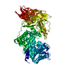
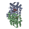
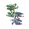
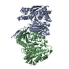
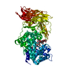
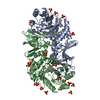
 Campylobacter jejuni (Campylobacter)
 X-RAY DIFFRACTION /
  Authors
 Citation
 Structure visualization
 Downloads & links
 Other downloads
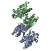


 PDBj
PDBj

 Assembly

 Mass: 62.068 Da / Num. of mol.: 2 / Source method: obtained synthetically / Formula: C2H6O2
 Mass: 96.063 Da / Num. of mol.: 4 / Source method: obtained synthetically / Formula: SO4
 Mass: 18.015 Da / Num. of mol.: 92 / Source method: isolated from a natural source / Formula: H2O
 Sample preparation
 / Beamline: 19-ID / Wavelength: 0.9772 Å
 Processing