 Movie
Movie Controller


+ Open data

- Basic information
| Entry | Database: PDB / ID: 1n7n | ||||||
|---|---|---|---|---|---|---|---|
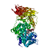
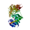
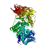
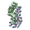
| Title | Streptococcus pneumoniae Hyaluronate Lyase W292A Mutant | ||||||
 Components Components | HYALURONIDASE | ||||||
 Keywords Keywords | LYASE / protein mutant | ||||||
| Function / homology |  Function and homology information Function and homology informationhyaluronate lyase / hyaluronate lyase activity / carbohydrate binding / carbohydrate metabolic process / extracellular region Similarity search - Function | ||||||
| Biological species |   Streptococcus pneumoniae (bacteria) Streptococcus pneumoniae (bacteria) | ||||||
| Method |  X-RAY DIFFRACTION / SYNCHROTRON / MOLECULAR REPLACEMENT / Resolution: 1.55 Å X-RAY DIFFRACTION / SYNCHROTRON / MOLECULAR REPLACEMENT / Resolution: 1.55 Å | ||||||
 Authors Authors | Nukui, M. / Taylor, K.B. / McPherson, D.T. / Shigenaga, M. / Jedrzejas, M.J. | ||||||
 Citation Citation | Journal: J.Biol.Chem. / Year: 2003 Title: The function of hydrophobic residues in the catalytic cleft of Streptococcus pneumoniae hyaluronate lyase. Kinetic characterization of mutant enzyme forms Authors: Nukui, M. / Taylor, K.B. / McPherson, D.T. / Shigenaga, M. / Jedrzejas, M.J. | ||||||
| History |
|
- Structure visualization
| Structure viewer | Molecule: MolmilJmol/JSmol |
|---|
- Downloads & links
-Download
| PDBx/mmCIF format | 1n7n.cif.gz | 172.8 KB | Display | PDBx/mmCIF format |
|---|---|---|---|---|
| PDB format | pdb1n7n.ent.gz | 132.4 KB | Display | PDB format |
| PDBx/mmJSON format | 1n7n.json.gz | Tree view | PDBx/mmJSON format | |
| Others |  Other downloads Other downloads |
-Validation report
| Arichive directory | https://data.pdbj.org/pub/pdb/validation_reports/n7/1n7nftp://data.pdbj.org/pub/pdb/validation_reports/n7/1n7n | HTTPS FTP |
|---|
-Related structure data
| Related structure data |  1n7oC  1n7pC  1n7qC  1n7rC  1lohS C: citing same article ( S: Starting model for refinement |
|---|---|
| Similar structure data |


-Links
 PDBj
PDBj


- Assembly
| Deposited unit | 
| ||||||||
|---|---|---|---|---|---|---|---|---|---|
| 1 |
| ||||||||
| Unit cell |
|
-Components
| #1: Protein | Mass: 82202.508 Da / Num. of mol.: 1 / Mutation: W292A mutant Source method: isolated from a genetically manipulated source Source: (gene. exp.) Streptococcus pneumoniae (bacteria) / Plasmid: pET20 / Production host: |
|---|---|
| #2: Water | ChemComp-HOH /  Mass: 18.015 Da / Num. of mol.: 732 / Source method: isolated from a natural source / Formula: H2O Mass: 18.015 Da / Num. of mol.: 732 / Source method: isolated from a natural source / Formula: H2O |
-Experimental details
-Experiment
| Experiment | Method: X-RAY DIFFRACTION / Number of used crystals: 2 |
|---|
- Sample preparation
| Crystal | Density Matthews: 2.43 Å3/Da / Density % sol: 49 % | ||||||||||||||||||||||||||||||||||||||||||
|---|---|---|---|---|---|---|---|---|---|---|---|---|---|---|---|---|---|---|---|---|---|---|---|---|---|---|---|---|---|---|---|---|---|---|---|---|---|---|---|---|---|---|---|
| Crystal grow | Temperature: 298 K / Method: vapor diffusion, hanging drop / pH: 6 Details: AMMONIUM SULFATE, SODIUM CACODYLATE, pH 6.0, VAPOR DIFFUSION, HANGING DROP, temperature 298K | ||||||||||||||||||||||||||||||||||||||||||
| Crystal grow | *PLUS | ||||||||||||||||||||||||||||||||||||||||||
| Components of the solutions | *PLUS
|
-Data collection
| Diffraction | Mean temperature: 100 K |
|---|---|
| Diffraction source | Source: SYNCHROTRON / Site: ALS  / Beamline: 5.0.1 / Wavelength: 1 Å / Beamline: 5.0.1 / Wavelength: 1 Å |
| Detector | Type: ADSC QUANTUM 4 / Detector: CCD / Date: Jan 10, 2002 |
| Radiation | Monochromator: SI crystal / Protocol: SINGLE WAVELENGTH / Monochromatic (M) / Laue (L): M / Scattering type: x-ray |
| Radiation wavelength | Wavelength: 1 Å / Relative weight: 1 |
| Reflection | Resolution: 1.55→50 Å / Num. obs: 125637 / % possible obs: 97.3 % / Observed criterion σ(F): 0 / Observed criterion σ(I): 0 / Redundancy: 5 % / Rmerge(I) obs: 0.058 / Rsym value: 0.058 |
| Reflection shell | Resolution: 1.55→1.61 Å / Rmerge(I) obs: 0.807 / % possible all: 87.5 |
| Reflection | *PLUS Lowest resolution: 50 Å / Num. obs: 125839 / Num. measured all: 636169 |
| Reflection shell | *PLUS % possible obs: 87.5 % |
- Processing
| Software |
| ||||||||||||||||
|---|---|---|---|---|---|---|---|---|---|---|---|---|---|---|---|---|---|
| Refinement | Method to determine structure: MOLECULAR REPLACEMENT Starting model: 1LOH Resolution: 1.55→20 Å / σ(F): 0 / σ(I): 0
| ||||||||||||||||
| Refinement step | Cycle: LAST / Resolution: 1.55→20 Å
| ||||||||||||||||
| LS refinement shell | Resolution: 1.55→1.61 Å
| ||||||||||||||||
| Refinement | *PLUS Lowest resolution: 50 Å / % reflection Rfree: 1 % / Rfactor obs: 0.18 / Rfactor Rwork: 0.18 | ||||||||||||||||
| Solvent computation | *PLUS | ||||||||||||||||
| Displacement parameters | *PLUS | ||||||||||||||||
| Refine LS restraints | *PLUS
| ||||||||||||||||
| LS refinement shell | *PLUS Rfactor Rwork: 0.28 |
