 Movie
Movie Controller

[English] 日本語
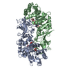
 Yorodumi
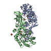
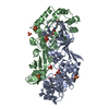
- PDB-3b8v: Crystal structure of Escherichia coli alaine racemase mutant E221K -
+ Open data

- Basic information
| Entry | Database: PDB / ID: 3b8v | ||||||
|---|---|---|---|---|---|---|---|
| Title | Crystal structure of Escherichia coli alaine racemase mutant E221K | ||||||
 Components Components | Alanine racemase | ||||||
 Keywords Keywords | ISOMERASE / alpha/beta barrel / Cell shape / Cell wall biogenesis/degradation / Peptidoglycan synthesis / Pyridoxal phosphate | ||||||
| Function / homology |  Function and homology information Function and homology informationalanine racemase / D-alanine biosynthetic process / alanine racemase activity / peptidoglycan biosynthetic process / cell wall organization / pyridoxal phosphate binding / regulation of cell shape / protein homodimerization activity / cytosol Similarity search - Function | ||||||
| Biological species |  | ||||||
| Method |  X-RAY DIFFRACTION / MOLECULAR REPLACEMENT / Resolution: 2.6 Å X-RAY DIFFRACTION / MOLECULAR REPLACEMENT / Resolution: 2.6 Å | ||||||
 Authors Authors | Wu, D. / Hu, T. / Zhang, L. / Jiang, H. / Shen, X. | ||||||
 Citation Citation | Journal: Protein Sci. / Year: 2008 Title: Residues Asp164 and Glu165 at the substrate entryway function potently in substrate orientation of alanine racemase from E. coli: Enzymatic characterization with crystal structure analysis Authors: Wu, D. / Hu, T. / Zhang, L. / Chen, J. / Du, J. / Ding, J. / Jiang, H. / Shen, X. | ||||||
| History |
|
- Structure visualization
| Structure viewer | Molecule: MolmilJmol/JSmol |
|---|
- Downloads & links
-Download
| PDBx/mmCIF format | 3b8v.cif.gz | 291.3 KB | Display | PDBx/mmCIF format |
|---|---|---|---|---|
| PDB format | pdb3b8v.ent.gz | 234.8 KB | Display | PDB format |
| PDBx/mmJSON format | 3b8v.json.gz | Tree view | PDBx/mmJSON format | |
| Others |  Other downloads Other downloads |
-Validation report
| Arichive directory | https://data.pdbj.org/pub/pdb/validation_reports/b8/3b8vftp://data.pdbj.org/pub/pdb/validation_reports/b8/3b8v | HTTPS FTP |
|---|
-Related structure data
| Related structure data |  2rjgSC 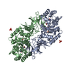 2rjhC  3b8tC  3b8uC  3b8wC S: Starting model for refinement C: citing same article ( |
|---|---|
| Similar structure data |
-Links
 PDBj
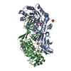
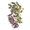
PDBj- Assembly
| Deposited unit | 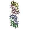
| ||||||||
|---|---|---|---|---|---|---|---|---|---|
| 1 | 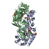
| ||||||||
| 2 | 
| ||||||||
| Unit cell |
|
-Components
| #1: Protein | Mass: 41416.301 Da / Num. of mol.: 4 / Mutation: E221K Source method: isolated from a genetically manipulated source Source: (gene. exp.) #2: Chemical | ChemComp-SO4 /   Mass: 96.063 Da / Num. of mol.: 11 / Source method: obtained synthetically / Formula: SO4 Mass: 96.063 Da / Num. of mol.: 11 / Source method: obtained synthetically / Formula: SO4#3: Chemical | ChemComp-PLP /   Mass: 247.142 Da / Num. of mol.: 4 / Source method: obtained synthetically / Formula: C8H10NO6P Mass: 247.142 Da / Num. of mol.: 4 / Source method: obtained synthetically / Formula: C8H10NO6P#4: Water | ChemComp-HOH / |  Mass: 18.015 Da / Num. of mol.: 489 / Source method: isolated from a natural source / Formula: H2O Mass: 18.015 Da / Num. of mol.: 489 / Source method: isolated from a natural source / Formula: H2O |
|---|
-Experimental details
-Experiment
| Experiment | Method: X-RAY DIFFRACTION / Number of used crystals: 1 |
|---|
- Sample preparation
| Crystal | Density Matthews: 3.13 Å3/Da / Density % sol: 60.72 % |
|---|---|
| Crystal grow | Temperature: 277 K / Method: vapor diffusion, hanging drop / pH: 6.6 Details: 0.1M MES, 1.6M ammonium sulfate, pH6.6, VAPOR DIFFUSION, HANGING DROP, temperature 277K |
-Data collection
| Diffraction | Mean temperature: 100 K | ||||||||||||||||||||||||||||||||||||||||||||||||||||||||||||||||||||||||||||||||||||||||
|---|---|---|---|---|---|---|---|---|---|---|---|---|---|---|---|---|---|---|---|---|---|---|---|---|---|---|---|---|---|---|---|---|---|---|---|---|---|---|---|---|---|---|---|---|---|---|---|---|---|---|---|---|---|---|---|---|---|---|---|---|---|---|---|---|---|---|---|---|---|---|---|---|---|---|---|---|---|---|---|---|---|---|---|---|---|---|---|---|---|
| Diffraction source | Source: ROTATING ANODE / Type: RIGAKU / Wavelength: 1.5418 Å | ||||||||||||||||||||||||||||||||||||||||||||||||||||||||||||||||||||||||||||||||||||||||
| Detector | Type: RIGAKU RAXIS IV / Detector: IMAGE PLATE / Date: Oct 18, 2007 | ||||||||||||||||||||||||||||||||||||||||||||||||||||||||||||||||||||||||||||||||||||||||
| Radiation | Protocol: SINGLE WAVELENGTH / Monochromatic (M) / Laue (L): M / Scattering type: x-ray | ||||||||||||||||||||||||||||||||||||||||||||||||||||||||||||||||||||||||||||||||||||||||
| Radiation wavelength | Wavelength: 1.5418 Å / Relative weight: 1 | ||||||||||||||||||||||||||||||||||||||||||||||||||||||||||||||||||||||||||||||||||||||||
| Reflection | Resolution: 2.6→164.399 Å / Num. obs: 62549 / % possible obs: 100 % / Redundancy: 9.3 % / Rmerge(I) obs: 0.141 / Rsym value: 0.141 / Net I/σ(I): 5.2 | ||||||||||||||||||||||||||||||||||||||||||||||||||||||||||||||||||||||||||||||||||||||||
| Reflection shell | Diffraction-ID: 1
|
- Processing
| Software |
| ||||||||||||||||||||||||||||
|---|---|---|---|---|---|---|---|---|---|---|---|---|---|---|---|---|---|---|---|---|---|---|---|---|---|---|---|---|---|
| Refinement | Method to determine structure: MOLECULAR REPLACEMENT Starting model: PDB ENTRY 2RJG Resolution: 2.6→15 Å / σ(F): 6 Details: This is a twinned structure, the detwin fraction is 0.400, and operator is 'h, -h-k, -l'.
| ||||||||||||||||||||||||||||
| Solvent computation | Bsol: 41.543 Å2 | ||||||||||||||||||||||||||||
| Displacement parameters | Biso mean: 22.421 Å2
| ||||||||||||||||||||||||||||
| Refinement step | Cycle: LAST / Resolution: 2.6→15 Å
| ||||||||||||||||||||||||||||
| Refine LS restraints |
| ||||||||||||||||||||||||||||
| Xplor file |
|
