 Movie
Movie Controller


+ Open data

- Basic information
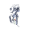
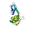
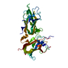
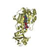
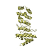
| Entry | Database: PDB / ID: 6dr3 | ||||||
|---|---|---|---|---|---|---|---|
| Title | Crystal structure of E. coli LpoA amino terminal domain | ||||||
 Components Components | Penicillin-binding protein activator LpoA | ||||||
 Keywords Keywords | BIOSYNTHETIC PROTEIN / TPR-like motifs OUTER MEMBRANE LIPOPROTEIN ACTIVATOR OF PBP1A PEPTIDOGLYCAN | ||||||
| Function / homology |  Function and homology information Function and homology informationperiplasmic side of cell outer membrane / hydrolase activity, hydrolyzing O-glycosyl compounds / peptidoglycan biosynthetic process / enzyme regulator activity / cell outer membrane / regulation of cell shape / periplasmic space / enzyme binding Similarity search - Function | ||||||
| Biological species |  | ||||||
| Method |  X-RAY DIFFRACTION / SYNCHROTRON / MOLECULAR REPLACEMENT / molecular replacement / Resolution: 2.101 Å X-RAY DIFFRACTION / SYNCHROTRON / MOLECULAR REPLACEMENT / molecular replacement / Resolution: 2.101 Å | ||||||
 Authors Authors | Kelley, A.C. / Saper, M.A. | ||||||
 Citation Citation | Journal: Acta Crystallogr.,Sect.F / Year: 2019 Title: Crystal structures of the amino-terminal domain of LpoA from Escherichia coli and Haemophilus influenzae. Authors: Kelley, A. / Vijayalakshmi, J. / Saper, M.A. #1: Journal: J. Biol. Chem. / Year: 2017Title: Structural analyses of the Haemophilus influenzae peptidoglycan synthase activator LpoA suggest multiple conformations in solution. Authors: Sathiyamoorthy, K. / Vijayalakshmi, J. / Tirupati, B. / Fan, L. / Saper, M.A. #2: Journal: Structure / Year: 2014Title: Elongated structure of the outer-membrane activator of peptidoglycan synthesis LpoA: implications for PBP1A stimulation. Authors: Jean, N.L. / Bougault, C.M. / Lodge, A. / Derouaux, A. / Callens, G. / Egan, A.J. / Ayala, I. / Lewis, R.J. / Vollmer, W. / Simorre, J.P. | ||||||
| History |
|
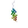
- Structure visualization
| Structure viewer | Molecule: MolmilJmol/JSmol |
|---|
- Downloads & links
-Download
| PDBx/mmCIF format | 6dr3.cif.gz | 144.6 KB | Display | PDBx/mmCIF format |
|---|---|---|---|---|
| PDB format | pdb6dr3.ent.gz | 115 KB | Display | PDB format |
| PDBx/mmJSON format | 6dr3.json.gz | Tree view | PDBx/mmJSON format | |
| Others |  Other downloads Other downloads |
-Validation report
| Arichive directory | https://data.pdbj.org/pub/pdb/validation_reports/dr/6dr3ftp://data.pdbj.org/pub/pdb/validation_reports/dr/6dr3 | HTTPS FTP |
|---|
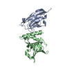
-Related structure data
| Related structure data | 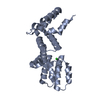 6dcjC 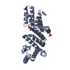 4p29S S: Starting model for refinement C: citing same article ( |
|---|---|
| Similar structure data |


-Links
 PDBj
PDBj- Assembly
| Deposited unit | 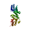
| ||||||||
|---|---|---|---|---|---|---|---|---|---|
| 1 |
| ||||||||
| Unit cell |
|
-Components
| #1: Protein | Mass: 25178.322 Da / Num. of mol.: 1 Source method: isolated from a genetically manipulated source Source: (gene. exp.) Strain: K12 / Gene: lpoA, yraM, b3147, JW3116 / Plasmid: pMCSG7-EcLpoA-N(31-252) Details (production host): Expresses of fusion of His6, TeV cleavage sequence, and EcLpoA-N(31-252) Cell line (production host): Origami 2(DE3) / Production host: |
|---|---|
| #2: Water | ChemComp-HOH /  Mass: 18.015 Da / Num. of mol.: 150 / Source method: isolated from a natural source / Formula: H2O Mass: 18.015 Da / Num. of mol.: 150 / Source method: isolated from a natural source / Formula: H2O |
-Experimental details
-Experiment
| Experiment | Method: X-RAY DIFFRACTION / Number of used crystals: 1 |
|---|
- Sample preparation
| Crystal | Density Matthews: 2.38 Å3/Da / Density % sol: 48.3 % / Description: needles with square cross-section |
|---|---|
| Crystal grow | Temperature: 298 K / Method: vapor diffusion, sitting drop / pH: 6.5 Details: Drops contained 0.5 ul protein and 0.5 ul precipitant and equilibrated against precipitant by vapor diffusion. Protein: 19 mg/ml in 50 mM NaCl, 1 mM dithiothreitol, 50 mM TrisHCl pH 7.5. ...Details: Drops contained 0.5 ul protein and 0.5 ul precipitant and equilibrated against precipitant by vapor diffusion. Protein: 19 mg/ml in 50 mM NaCl, 1 mM dithiothreitol, 50 mM TrisHCl pH 7.5. Precipitant contained: 0.03 M magnesium chloride hexahydrate, 0.03 M calcium chloride dihydrate, 5% glycerol, 0.1 M Buffer System 1, pH 6.5, 10% PEG 20,000, 17% PEG 550 MME Buffer System 1: 30 mL MES (pH 3.11) was titrated with 24.1 mL Imidazole (pH 10.23) to a final pH of 6.5 |
-Data collection
| Diffraction | Mean temperature: 140 K | |||||||||||||||||||||||||||||||||||||||||||||||||||||||||||||||||||||||||||||||||||||||||||||||||||
|---|---|---|---|---|---|---|---|---|---|---|---|---|---|---|---|---|---|---|---|---|---|---|---|---|---|---|---|---|---|---|---|---|---|---|---|---|---|---|---|---|---|---|---|---|---|---|---|---|---|---|---|---|---|---|---|---|---|---|---|---|---|---|---|---|---|---|---|---|---|---|---|---|---|---|---|---|---|---|---|---|---|---|---|---|---|---|---|---|---|---|---|---|---|---|---|---|---|---|---|---|
| Diffraction source | Source: SYNCHROTRON / Site: APS  / Beamline: 21-ID-D / Wavelength: 0.97717 Å / Beamline: 21-ID-D / Wavelength: 0.97717 Å | |||||||||||||||||||||||||||||||||||||||||||||||||||||||||||||||||||||||||||||||||||||||||||||||||||
| Detector | Type: DECTRIS EIGER X 9M / Detector: PIXEL / Date: Aug 2, 2017 / Details: mirrors | |||||||||||||||||||||||||||||||||||||||||||||||||||||||||||||||||||||||||||||||||||||||||||||||||||
| Radiation | Monochromator: Kohzu / Protocol: SINGLE WAVELENGTH / Monochromatic (M) / Laue (L): M / Scattering type: x-ray | |||||||||||||||||||||||||||||||||||||||||||||||||||||||||||||||||||||||||||||||||||||||||||||||||||
| Radiation wavelength | Wavelength: 0.97717 Å / Relative weight: 1 | |||||||||||||||||||||||||||||||||||||||||||||||||||||||||||||||||||||||||||||||||||||||||||||||||||
| Reflection | Resolution: 2.1→30 Å / Num. obs: 14791 / % possible obs: 99.8 % / Redundancy: 10.9 % / Biso Wilson estimate: 27.23 Å2 / Rmerge(I) obs: 0.104 / Rpim(I) all: 0.032 / Rrim(I) all: 0.109 / Χ2: 0.959 / Net I/σ(I): 6.7 / Num. measured all: 160798 | |||||||||||||||||||||||||||||||||||||||||||||||||||||||||||||||||||||||||||||||||||||||||||||||||||
| Reflection shell | Diffraction-ID: 1
|
-Phasing
| Phasing | Method: molecular replacement | |||||||||
|---|---|---|---|---|---|---|---|---|---|---|
| Phasing MR |
|
- Processing
| Software |
| |||||||||||||||||||||||||||||||||||||||||||||||||||||||||||||||||||||||||||||
|---|---|---|---|---|---|---|---|---|---|---|---|---|---|---|---|---|---|---|---|---|---|---|---|---|---|---|---|---|---|---|---|---|---|---|---|---|---|---|---|---|---|---|---|---|---|---|---|---|---|---|---|---|---|---|---|---|---|---|---|---|---|---|---|---|---|---|---|---|---|---|---|---|---|---|---|---|---|---|
| Refinement | Method to determine structure: MOLECULAR REPLACEMENT Starting model: 4P29 Resolution: 2.101→22.697 Å / SU ML: 0.19 / Cross valid method: THROUGHOUT / σ(F): 1.47 / Phase error: 20.19
| |||||||||||||||||||||||||||||||||||||||||||||||||||||||||||||||||||||||||||||
| Solvent computation | Shrinkage radii: 0.9 Å / VDW probe radii: 1.11 Å | |||||||||||||||||||||||||||||||||||||||||||||||||||||||||||||||||||||||||||||
| Displacement parameters | Biso max: 158.63 Å2 / Biso mean: 42.4992 Å2 / Biso min: 14.45 Å2 | |||||||||||||||||||||||||||||||||||||||||||||||||||||||||||||||||||||||||||||
| Refinement step | Cycle: final / Resolution: 2.101→22.697 Å
| |||||||||||||||||||||||||||||||||||||||||||||||||||||||||||||||||||||||||||||
| Refine LS restraints |
| |||||||||||||||||||||||||||||||||||||||||||||||||||||||||||||||||||||||||||||
| LS refinement shell | Refine-ID: X-RAY DIFFRACTION / Rfactor Rfree error: 0 / Total num. of bins used: 10
| |||||||||||||||||||||||||||||||||||||||||||||||||||||||||||||||||||||||||||||
| Refinement TLS params. | Method: refined / Refine-ID: X-RAY DIFFRACTION
| |||||||||||||||||||||||||||||||||||||||||||||||||||||||||||||||||||||||||||||
| Refinement TLS group |
|
