 Movie
Movie Controller


+ Open data

- Basic information
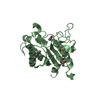
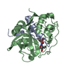
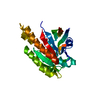
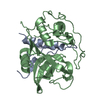
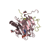
| Entry | Database: PDB / ID: 6ahs | ||||||
|---|---|---|---|---|---|---|---|
| Title | Mouse Kallikrein 7 in complex with imidazolinylindole derivative | ||||||
 Components Components | Kallikrein-7 | ||||||
 Keywords Keywords | HYDROLASE / protease | ||||||
| Function / homology |  Function and homology informationstratum corneum chymotryptic enzyme / positive regulation of antibacterial peptide production / epidermal lamellar body / Degradation of the extracellular matrix / cornified envelope / secretory granule / peptidase activity / serine-type endopeptidase activity / proteolysis / extracellular space Function and homology informationstratum corneum chymotryptic enzyme / positive regulation of antibacterial peptide production / epidermal lamellar body / Degradation of the extracellular matrix / cornified envelope / secretory granule / peptidase activity / serine-type endopeptidase activity / proteolysis / extracellular spaceSimilarity search - Function | ||||||
| Biological species |  Mus musculus (house mouse) Mus musculus (house mouse) | ||||||
| Method | X-RAY DIFFRACTION / MOLECULAR REPLACEMENT / Resolution: 1.75 Å | ||||||
 Authors Authors | Sugawara, H. | ||||||
 Citation Citation | Journal: Bioorg. Med. Chem. Lett. / Year: 2019 Title: Discovery and structure-activity relationship of imidazolinylindole derivatives as kallikrein 7 inhibitors. Authors: Murafuji, H. / Muto, T. / Goto, M. / Imajo, S. / Sugawara, H. / Oyama, Y. / Minamitsuji, Y. / Miyazaki, S. / Murai, K. / Fujioka, H. #1: Journal: Bioorg. Med. Chem. Lett. / Year: 2017Title: Discovery and structure-activity relationship study of 1,3,6-trisubstituted 1,4-diazepane-7-ones as novel human kallikrein 7 inhibitors. Authors: Murafuji, H. / Sakai, H. / Goto, M. / Imajo, S. / Sugawara, H. / Muto, T. #2: Journal: Bioorg. Med. Chem. Lett. / Year: 2018 Title: Structure-based drug design of 1,3,6-trisubstituted 1,4-diazepan-7-ones as selective human kallikrein 7 inhibitors. Authors: Murafuji, H. / Sakai, H. / Goto, M. / Oyama, Y. / Imajo, S. / Sugawara, H. / Tomoo, T. / Muto, T. #3: Journal: Bioorg. Med. Chem. / Year: 2018Title: Structure-based drug design to overcome species differences in kallikrein 7 inhibition of 1,3,6-trisubstituted 1,4-diazepan-7-ones. Authors: Murafuji, H. / Sugawara, H. / Goto, M. / Oyama, Y. / Sakai, H. / Imajo, S. / Tomoo, T. / Muto, T. | ||||||
| History |
|
- Structure visualization
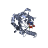
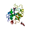
| Structure viewer | Molecule: MolmilJmol/JSmol |
|---|
- Downloads & links
-Download
| PDBx/mmCIF format | 6ahs.cif.gz | 104.7 KB | Display | PDBx/mmCIF format |
|---|---|---|---|---|
| PDB format | pdb6ahs.ent.gz | 78.3 KB | Display | PDB format |
| PDBx/mmJSON format | 6ahs.json.gz | Tree view | PDBx/mmJSON format | |
| Others |  Other downloads Other downloads |
-Validation report
| Arichive directory | https://data.pdbj.org/pub/pdb/validation_reports/ah/6ahsftp://data.pdbj.org/pub/pdb/validation_reports/ah/6ahs | HTTPS FTP |
|---|
-Related structure data
| Related structure data |  5zfhS S: Starting model for refinement |
|---|---|
| Similar structure data |


-Links
 PDBj
PDBj
- Assembly
| Deposited unit | 
| |||||||||
|---|---|---|---|---|---|---|---|---|---|---|
| 1 |
| |||||||||
| Unit cell |
| |||||||||
| Components on special symmetry positions |
|
-Components
| #1: Protein | / Serine protease 6 / Stratum corneum chymotryptic enzyme / Thymopsin Mass: 24637.334 Da / Num. of mol.: 1 Source method: isolated from a genetically manipulated source Source: (gene. exp.) Mus musculus (house mouse) / Gene: Klk7, Prss6, Scce / Production host:  Escherichia coli (E. coli) Escherichia coli (E. coli)References: UniProt: Q91VE3, stratum corneum chymotryptic enzyme | ||
|---|---|---|---|
| #2: Chemical | ChemComp-9YO /   Mass: 387.883 Da / Num. of mol.: 1 / Source method: obtained synthetically / Formula: C19H18ClN3O2S / Feature type: SUBJECT OF INVESTIGATION Mass: 387.883 Da / Num. of mol.: 1 / Source method: obtained synthetically / Formula: C19H18ClN3O2S / Feature type: SUBJECT OF INVESTIGATION | ||
| #3: Chemical | ChemComp-TRS / Tris  Mass: 122.143 Da / Num. of mol.: 1 / Source method: obtained synthetically / Formula: C4H12NO3 / Comment: pH buffer*YM Mass: 122.143 Da / Num. of mol.: 1 / Source method: obtained synthetically / Formula: C4H12NO3 / Comment: pH buffer*YM | ||
| #4: Chemical | Chloride  Mass: 35.453 Da / Num. of mol.: 2 / Source method: obtained synthetically / Formula: Cl Mass: 35.453 Da / Num. of mol.: 2 / Source method: obtained synthetically / Formula: Cl#5: Water | ChemComp-HOH / | Water Mass: 18.015 Da / Num. of mol.: 142 / Source method: isolated from a natural source / Formula: H2O Mass: 18.015 Da / Num. of mol.: 142 / Source method: isolated from a natural source / Formula: H2O |
-Experimental details
-Experiment
| Experiment | Method: X-RAY DIFFRACTION / Number of used crystals: 1 |
|---|
- Sample preparation
| Crystal | Density Matthews: 2.54 Å3/Da / Density % sol: 51.59 % |
|---|---|
| Crystal grow | Temperature: 293 K / Method: vapor diffusion, sitting drop / pH: 8 / Details: PEG 8000, PEG 400, magnesium chloride, Tris/HCl |
-Data collection
| Diffraction | Mean temperature: 100 K |
|---|---|
| Diffraction source | Source: ROTATING ANODE / Type: RIGAKU MICROMAX-007 / Wavelength: 1.5418 Å |
| Detector | Type: RIGAKU RAXIS IV++ / Detector: IMAGE PLATE / Date: Jul 10, 2012 |
| Radiation | Protocol: SINGLE WAVELENGTH / Monochromatic (M) / Laue (L): M / Scattering type: x-ray |
| Radiation wavelength | Wavelength: 1.5418 Å / Relative weight: 1 |
| Reflection | Resolution: 1.75→100 Å / Num. obs: 26249 / % possible obs: 99.1 % / Redundancy: 13.2 % / Net I/σ(I): 17.6 |
| Reflection shell | Resolution: 1.75→1.78 Å |
- Processing
| Software |
| ||||||||||||||||||||||||||||||||||||||||||||||||||||||||||||||||||||||||||||||||||||||||||||||||||||||||||||||||||||||||||||||||||||||||||||||||||||||||||||||||||||||||||||||||||||||
|---|---|---|---|---|---|---|---|---|---|---|---|---|---|---|---|---|---|---|---|---|---|---|---|---|---|---|---|---|---|---|---|---|---|---|---|---|---|---|---|---|---|---|---|---|---|---|---|---|---|---|---|---|---|---|---|---|---|---|---|---|---|---|---|---|---|---|---|---|---|---|---|---|---|---|---|---|---|---|---|---|---|---|---|---|---|---|---|---|---|---|---|---|---|---|---|---|---|---|---|---|---|---|---|---|---|---|---|---|---|---|---|---|---|---|---|---|---|---|---|---|---|---|---|---|---|---|---|---|---|---|---|---|---|---|---|---|---|---|---|---|---|---|---|---|---|---|---|---|---|---|---|---|---|---|---|---|---|---|---|---|---|---|---|---|---|---|---|---|---|---|---|---|---|---|---|---|---|---|---|---|---|---|---|
| Refinement | Method to determine structure: MOLECULAR REPLACEMENT Starting model: 5ZFH Resolution: 1.75→30.29 Å / Cor.coef. Fo:Fc: 0.966 / Cor.coef. Fo:Fc free: 0.945 / SU B: 4.055 / SU ML: 0.063 / Cross valid method: THROUGHOUT / ESU R: 0.022 / ESU R Free: 0.023 / Details: HYDROGENS HAVE BEEN ADDED IN THE RIDING POSITIONS
| ||||||||||||||||||||||||||||||||||||||||||||||||||||||||||||||||||||||||||||||||||||||||||||||||||||||||||||||||||||||||||||||||||||||||||||||||||||||||||||||||||||||||||||||||||||||
| Solvent computation | Ion probe radii: 0.8 Å / Shrinkage radii: 0.8 Å / VDW probe radii: 1.2 Å | ||||||||||||||||||||||||||||||||||||||||||||||||||||||||||||||||||||||||||||||||||||||||||||||||||||||||||||||||||||||||||||||||||||||||||||||||||||||||||||||||||||||||||||||||||||||
| Displacement parameters | Biso mean: 29.665 Å2
| ||||||||||||||||||||||||||||||||||||||||||||||||||||||||||||||||||||||||||||||||||||||||||||||||||||||||||||||||||||||||||||||||||||||||||||||||||||||||||||||||||||||||||||||||||||||
| Refinement step | Cycle: 1 / Resolution: 1.75→30.29 Å
| ||||||||||||||||||||||||||||||||||||||||||||||||||||||||||||||||||||||||||||||||||||||||||||||||||||||||||||||||||||||||||||||||||||||||||||||||||||||||||||||||||||||||||||||||||||||
| Refine LS restraints |
|