 Movie
Movie Controller

[English] 日本語
 Yorodumi
- PDB-1csb: Crystal structure of cathepsin b inhibited with CA030 at 2.1 angs... -
+ Open data

- Basic information
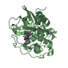
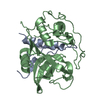
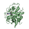
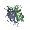
| Entry | Database: PDB / ID: 1csb | ||||||
|---|---|---|---|---|---|---|---|
| Title | Crystal structure of cathepsin b inhibited with CA030 at 2.1 angstroms resolution: A basis for the design of specific epoxysuccinyl inhibitors | ||||||
 Components Components |
| ||||||
 Keywords Keywords | HYDROLASE/HYDROLASE INHIBITOR / PAPAIN-LIKE LYSOSOMAL DICARBOXY-PEPTIDASE / HYDROLASE-HYDROLASE INHIBITOR complex | ||||||
| Function / homology |  Function and homology information Function and homology informationcathepsin B / peptidase inhibitor complex / endolysosome lumen / thyroid hormone generation / cellular response to thyroid hormone stimulus / Trafficking and processing of endosomal TLR / proteoglycan binding / Assembly of collagen fibrils and other multimeric structures / Collagen degradation / decidualization ...cathepsin B / peptidase inhibitor complex / endolysosome lumen / thyroid hormone generation / cellular response to thyroid hormone stimulus / Trafficking and processing of endosomal TLR / proteoglycan binding / Assembly of collagen fibrils and other multimeric structures / Collagen degradation / decidualization / collagen catabolic process / collagen binding / epithelial cell differentiation / cysteine-type peptidase activity / MHC class II antigen presentation / : / Degradation of CDH1 / melanosome / peptidase activity / extracellular matrix / regulation of apoptotic process / ficolin-1-rich granule lumen / lysosome / apical plasma membrane / external side of plasma membrane / cysteine-type endopeptidase activity / Neutrophil degranulation / symbiont entry into host cell / perinuclear region of cytoplasm / proteolysis / : / extracellular exosome / extracellular region Similarity search - Function | ||||||
| Biological species |  Homo sapiens (human) Homo sapiens (human) | ||||||
| Method |  X-RAY DIFFRACTION / Resolution: 2 Å X-RAY DIFFRACTION / Resolution: 2 Å | ||||||
 Authors Authors | Turk, D. / Bode, W. | ||||||
 Citation Citation | Journal: Biochemistry / Year: 1995 Title: Crystal structure of cathepsin B inhibited with CA030 at 2.0-A resolution: A basis for the design of specific epoxysuccinyl inhibitors. Authors: Turk, D. / Podobnik, M. / Popovic, T. / Katunuma, N. / Bode, W. / Huber, R. / Turk, V. #1: Journal: Embo J. / Year: 1991Title: The Refined 2.15 Angstrom X-Ray Crystal Structure of Human Liver Cathepsin B: The Structural Basis for its Specificity Authors: Musil, D. / Zucic, D. / Turk, D. / Engh, R.A. / Mayr, I. / Huber, R. / Popovic, T. / Turk, V. / Towatari, T. / Katunuma, N. / Bode, W. | ||||||
| History |
|
- Structure visualization
| Structure viewer | Molecule: MolmilJmol/JSmol |
|---|
- Downloads & links
-Download
| PDBx/mmCIF format | 1csb.cif.gz | 120.9 KB | Display | PDBx/mmCIF format |
|---|---|---|---|---|
| PDB format | pdb1csb.ent.gz | 93 KB | Display | PDB format |
| PDBx/mmJSON format | 1csb.json.gz | Tree view | PDBx/mmJSON format | |
| Others |  Other downloads Other downloads |
-Validation report
| Arichive directory | https://data.pdbj.org/pub/pdb/validation_reports/cs/1csbftp://data.pdbj.org/pub/pdb/validation_reports/cs/1csb | HTTPS FTP |
|---|
-Related structure data
| Similar structure data |
|---|


-Links
 PDBj
PDBj


- Assembly
| Deposited unit | 
| ||||||||
|---|---|---|---|---|---|---|---|---|---|
| 1 | 
| ||||||||
| 2 | 
| ||||||||
| Unit cell |
| ||||||||
| Atom site foot note | 1: CIS PROLINE - PRO B 138 / 2: CIS PROLINE - PRO E 138 | ||||||||
| Noncrystallographic symmetry (NCS) | NCS oper: (Code: given Matrix: (0.018575, -0.006577, -0.999806), Vector: Details | MTRIX THE TRANSFORMATIONS PRESENTED ON MTRIX RECORDS BELOW DESCRIBE NON-CRYSTALLOGRAPHIC RELATIONSHIPS AMONG THE VARIOUS DOMAINS IN THIS ENTRY. APPLYING THE APPROPRIATE MTRIX TRANSFORMATION TO THE RESIDUES LISTED FIRST WILL YIELD APPROXIMATE COORDINATES FOR THE RESIDUES LISTED SECOND. APPLIED TO TRANSFORMED TO MTRIX RESIDUES RESIDUES RMSD M1 D 1 .. D 47 A 1 .. A 47 0.432 M1 E 50 .. E 254 B 50 .. B 254 0.562 M1 F 3 .. F 4 C 3 .. C 4 0.456 | |
-Components
| #1: Protein/peptide | Mass: 5213.840 Da / Num. of mol.: 2 / Source method: isolated from a natural source / Source: (natural) Homo sapiens (human) / Organ: LIVER / Organelle: LYSOSOME / References: UniProt: P07858, cathepsin B#2: Antibody | Mass: 22437.910 Da / Num. of mol.: 2 / Source method: isolated from a natural source / Source: (natural) Homo sapiens (human) / Organ: LIVER / Organelle: LYSOSOME / References: UniProt: P07858, cathepsin B#3: Chemical |   Type: Peptide-like / Class: Inhibitor / Mass: 372.413 Da / Num. of mol.: 2 / Source method: obtained synthetically / Formula: C17H28N2O7 / References: CA030, ethyl ester of epoxysuccinyl-Ile-Pro-OH Type: Peptide-like / Class: Inhibitor / Mass: 372.413 Da / Num. of mol.: 2 / Source method: obtained synthetically / Formula: C17H28N2O7 / References: CA030, ethyl ester of epoxysuccinyl-Ile-Pro-OH#4: Water | ChemComp-HOH / |  Mass: 18.015 Da / Num. of mol.: 542 / Source method: isolated from a natural source / Formula: H2O Mass: 18.015 Da / Num. of mol.: 542 / Source method: isolated from a natural source / Formula: H2OHas protein modification | Y | |
|---|
-Experimental details
-Experiment
| Experiment | Method: X-RAY DIFFRACTION |
|---|
- Sample preparation
| Crystal | Density Matthews: 2.2 Å3/Da / Density % sol: 44.07 % | ||||||||||||||||||||
|---|---|---|---|---|---|---|---|---|---|---|---|---|---|---|---|---|---|---|---|---|---|
| Crystal grow | *PLUS Temperature: 21 ℃ / pH: 5 / Method: vapor diffusion, hanging drop | ||||||||||||||||||||
| Components of the solutions | *PLUS
|
-Data collection
| Diffraction source | Wavelength: 1.5418 Å |
|---|---|
| Detector | Type: MARRESEARCH / Detector: IMAGE PLATE / Date: Nov 1, 1993 |
| Radiation | Monochromatic (M) / Laue (L): M / Scattering type: x-ray |
| Radiation wavelength | Wavelength: 1.5418 Å / Relative weight: 1 |
| Reflection | Num. obs: 51421 / % possible obs: 79.1 % / Observed criterion σ(I): 2 / Rmerge(I) obs: 0.098 |
| Reflection | *PLUS Highest resolution: 2 Å / Rmerge(I) obs: 0.098 |
| Reflection shell | *PLUS Highest resolution: 2 Å / Lowest resolution: 2.05 Å / % possible obs: 64 % |
- Processing
| Software |
| ||||||||||||||||||||||||||||||||||||||||||||||||||||||||||||
|---|---|---|---|---|---|---|---|---|---|---|---|---|---|---|---|---|---|---|---|---|---|---|---|---|---|---|---|---|---|---|---|---|---|---|---|---|---|---|---|---|---|---|---|---|---|---|---|---|---|---|---|---|---|---|---|---|---|---|---|---|---|
| Refinement | Resolution: 2→8 Å / σ(F): 2 Details: THE MINIMAL ATOMIC TEMPERATURE FACTOR WAS SET TO 5.
| ||||||||||||||||||||||||||||||||||||||||||||||||||||||||||||
| Refinement step | Cycle: LAST / Resolution: 2→8 Å
| ||||||||||||||||||||||||||||||||||||||||||||||||||||||||||||
| Refine LS restraints |
| ||||||||||||||||||||||||||||||||||||||||||||||||||||||||||||
| Software | *PLUS Name: X-PLOR / Classification: refinement | ||||||||||||||||||||||||||||||||||||||||||||||||||||||||||||
| Refinement | *PLUS | ||||||||||||||||||||||||||||||||||||||||||||||||||||||||||||
| Solvent computation | *PLUS | ||||||||||||||||||||||||||||||||||||||||||||||||||||||||||||
| Displacement parameters | *PLUS | ||||||||||||||||||||||||||||||||||||||||||||||||||||||||||||
| Refine LS restraints | *PLUS Type: x_bond_d / Dev ideal: 0.011 |
