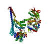
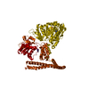
- PDB-5zui: Crystal Structure of HSP104 from Chaetomium thermophilum -
+
Open data
ID or keywords:
Loading...
-
Basic information
Entry
Database: PDB / ID: 5zui
Title
Crystal Structure of HSP104 from Chaetomium thermophilum
Components
Heat Shock Protein 104
Keywords
CHAPERONE / PROTEIN DISAGGREGASE / ATPASE / TWO-RING AAA PROTEIN / HELICAL FILAMENT
Function / homology
Function and homology information
cellular heat acclimation / protein unfolding / unfolded protein binding / protein-folding chaperone binding / protein refolding / ATP hydrolysis activity / ATP binding / cytosol Similarity search - Function
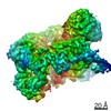
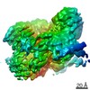
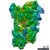
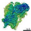
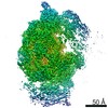
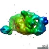
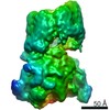
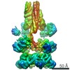
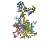
Journal: Structure / Year: 2021 Title: Split conformation of Chaetomium thermophilum Hsp104 disaggregase. Authors: Yosuke Inoue / Yuya Hanazono / Kentaro Noi / Akihiro Kawamoto / Masato Kimatsuka / Ryuhei Harada / Kazuki Takeda / Ryoichi Kita / Natsuki Iwamasa / Kyoka Shibata / Keiichi Noguchi / Yasuteru ...Authors: Yosuke Inoue / Yuya Hanazono / Kentaro Noi / Akihiro Kawamoto / Masato Kimatsuka / Ryuhei Harada / Kazuki Takeda / Ryoichi Kita / Natsuki Iwamasa / Kyoka Shibata / Keiichi Noguchi / Yasuteru Shigeta / Keiichi Namba / Teru Ogura / Kunio Miki / Kyosuke Shinohara / Masafumi Yohda / Abstract: Hsp104 and its bacterial homolog ClpB form hexameric ring structures and mediate protein disaggregation. The disaggregated polypeptide is thought to thread through the central channel of the ring. ...Hsp104 and its bacterial homolog ClpB form hexameric ring structures and mediate protein disaggregation. The disaggregated polypeptide is thought to thread through the central channel of the ring. However, the dynamic behavior of Hsp104 during disaggregation remains unclear. Here, we reported the stochastic conformational dynamics and a split conformation of Hsp104 disaggregase from Chaetomium thermophilum (CtHsp104) in the presence of ADP by X-ray crystallography, cryo-electron microscopy (EM), and high-speed atomic force microscopy (AFM). ADP-bound CtHsp104 assembles into a 6 left-handed spiral filament in the crystal structure at a resolution of 2.7 Å. The unit of the filament is a hexamer of the split spiral structure. In the cryo-EM images, staggered and split hexameric rings were observed. Further, high-speed AFM observations showed that a substrate addition enhanced the conformational change and increased the split structure's frequency. Our data suggest that split conformation is an off-pathway state of CtHsp104 during disaggregation.
History
Deposition
May 7, 2018
Deposition site: PDBJ / Processing site: PDBJ
Revision 1.0
Jun 19, 2019
Provider: repository / Type: Initial release
Revision 1.1
Oct 16, 2019
Group: Data collection / Category: reflns / Item: _reflns.pdbx_CC_half
In the structure databanks used in Yorodumi, some data are registered as the other names, "COVID-19 virus" and "2019-nCoV". Here are the details of the virus and the list of structure data.
Jan 31, 2019. EMDB accession codes are about to change! (news from PDBe EMDB page)
EMDB accession codes are about to change! (news from PDBe EMDB page)
The allocation of 4 digits for EMDB accession codes will soon come to an end. Whilst these codes will remain in use, new EMDB accession codes will include an additional digit and will expand incrementally as the available range of codes is exhausted. The current 4-digit format prefixed with “EMD-” (i.e. EMD-XXXX) will advance to a 5-digit format (i.e. EMD-XXXXX), and so on. It is currently estimated that the 4-digit codes will be depleted around Spring 2019, at which point the 5-digit format will come into force.
The EM Navigator/Yorodumi systems omit the EMD- prefix.
Related info.:Q: What is EMD? / ID/Accession-code notation in Yorodumi/EM Navigator
Yorodumi is a browser for structure data from EMDB, PDB, SASBDB, etc.
This page is also the successor to EM Navigator detail page, and also detail information page/front-end page for Omokage search.
The word "yorodu" (or yorozu) is an old Japanese word meaning "ten thousand". "mi" (miru) is to see.
Related info.:EMDB / PDB / SASBDB / Comparison of 3 databanks / Yorodumi Search / Aug 31, 2016. New EM Navigator & Yorodumi / Yorodumi Papers / Jmol/JSmol / Function and homology information / Changes in new EM Navigator and Yorodumi
 Movie
Movie Controller


 Open data
 Basic information
 Components
 Keywords
 Function and homology information
 Chaetomium thermophilum (fungus)
 X-RAY DIFFRACTION /
  Authors
 Citation
 Structure visualization
 Downloads & links
 Other downloads
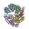


 PDBj
PDBj


 Assembly
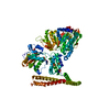


 Mass: 427.201 Da / Num. of mol.: 2 / Source method: obtained synthetically / Formula: C10H15N5O10P2 / Comment: ADP, energy-carrying molecule*YM
 Mass: 96.063 Da / Num. of mol.: 6 / Source method: obtained synthetically / Formula: SO4
 Mass: 18.015 Da / Num. of mol.: 5 / Source method: isolated from a natural source / Formula: H2O
 Sample preparation
 Processing