 Movie
Movie Controller

[English] 日本語
 Yorodumi
- PDB-5no4: RsgA-GDPNP bound to the 30S ribosomal subunit (RsgA assembly inte... -
+ Open data

- Basic information
| Entry | Database: PDB / ID: 5no4 | ||||||
|---|---|---|---|---|---|---|---|
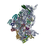
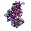
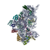
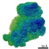
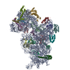
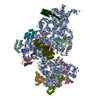
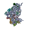
| Title | RsgA-GDPNP bound to the 30S ribosomal subunit (RsgA assembly intermediate with uS3) | ||||||
 Components Components |
| ||||||
 Keywords Keywords | RIBOSOME | ||||||
| Function / homology |  Function and homology information Function and homology informationguanosine tetraphosphate binding / transcription antitermination factor activity, RNA binding / ornithine decarboxylase inhibitor activity / misfolded RNA binding / Group I intron splicing / RNA folding / Hydrolases; Acting on acid anhydrides; In phosphorus-containing anhydrides / four-way junction DNA binding / negative regulation of translational initiation / regulation of mRNA stability ...guanosine tetraphosphate binding / transcription antitermination factor activity, RNA binding / ornithine decarboxylase inhibitor activity / misfolded RNA binding / Group I intron splicing / RNA folding / Hydrolases; Acting on acid anhydrides; In phosphorus-containing anhydrides / four-way junction DNA binding / negative regulation of translational initiation / regulation of mRNA stability / mRNA regulatory element binding translation repressor activity / positive regulation of RNA splicing / regulation of DNA-templated transcription elongation / transcription elongation factor complex / transcription antitermination / DNA endonuclease activity / DNA-templated transcription termination / maintenance of translational fidelity / mRNA 5'-UTR binding / GDP binding / regulation of translation / ribosome biogenesis / ribosomal small subunit assembly / ribosomal small subunit biogenesis / small ribosomal subunit / small ribosomal subunit rRNA binding / cytosolic small ribosomal subunit / cytoplasmic translation / tRNA binding / negative regulation of translation / rRNA binding / structural constituent of ribosome / ribosome / translation / hydrolase activity / response to antibiotic / GTPase activity / mRNA binding / GTP binding / RNA binding / zinc ion binding / metal ion binding / membrane / cytoplasm / cytosol Similarity search - Function | ||||||
| Biological species |  | ||||||
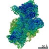
| Method | ELECTRON MICROSCOPY / single particle reconstruction / cryo EM / Resolution: 5.16 Å | ||||||
 Authors Authors | Lopez-Alonso, J.P. / Kaminishi, T. / Kikuchi, T. / Hirata, Y. / Iturrioz, I. / Dhimole, N. / Schedlbauer, A. / Hase, Y. / Goto, S. / Kurita, D. ...Lopez-Alonso, J.P. / Kaminishi, T. / Kikuchi, T. / Hirata, Y. / Iturrioz, I. / Dhimole, N. / Schedlbauer, A. / Hase, Y. / Goto, S. / Kurita, D. / Muto, A. / Zhou, S. / Naoe, C. / Mills, D.J. / Gil-Carton, D. / Takemoto, C. / Himeno, H. / Fucini, P. / Connell, S.R. | ||||||
 Citation Citation | Journal: Nucleic Acids Res / Year: 2017 Title: RsgA couples the maturation state of the 30S ribosomal decoding center to activation of its GTPase pocket. Authors: Jorge Pedro López-Alonso / Tatsuya Kaminishi / Takeshi Kikuchi / Yuya Hirata / Idoia Iturrioz / Neha Dhimole / Andreas Schedlbauer / Yoichi Hase / Simon Goto / Daisuke Kurita / Akira Muto / ...Authors: Jorge Pedro López-Alonso / Tatsuya Kaminishi / Takeshi Kikuchi / Yuya Hirata / Idoia Iturrioz / Neha Dhimole / Andreas Schedlbauer / Yoichi Hase / Simon Goto / Daisuke Kurita / Akira Muto / Shu Zhou / Chieko Naoe / Deryck J Mills / David Gil-Carton / Chie Takemoto / Hyouta Himeno / Paola Fucini / Sean R Connell /    Abstract: During 30S ribosomal subunit biogenesis, assembly factors are believed to prevent accumulation of misfolded intermediate states of low free energy that slowly convert into mature 30S subunits, ...During 30S ribosomal subunit biogenesis, assembly factors are believed to prevent accumulation of misfolded intermediate states of low free energy that slowly convert into mature 30S subunits, namely, kinetically trapped particles. Among the assembly factors, the circularly permuted GTPase, RsgA, plays a crucial role in the maturation of the 30S decoding center. Here, directed hydroxyl radical probing and single particle cryo-EM are employed to elucidate RsgA΄s mechanism of action. Our results show that RsgA destabilizes the 30S structure, including late binding r-proteins, providing a structural basis for avoiding kinetically trapped assembly intermediates. Moreover, RsgA exploits its distinct GTPase pocket and specific interactions with the 30S to coordinate GTPase activation with the maturation state of the 30S subunit. This coordination validates the architecture of the decoding center and facilitates the timely release of RsgA to control the progression of 30S biogenesis. | ||||||
| History |
|
- Structure visualization
| Movie |
Movie viewer |
|---|---|
| Structure viewer | Molecule: MolmilJmol/JSmol |
- Downloads & links
-Download
| PDBx/mmCIF format | 5no4.cif.gz | 1.1 MB | Display | PDBx/mmCIF format |
|---|---|---|---|---|
| PDB format | pdb5no4.ent.gz | 889.5 KB | Display | PDB format |
| PDBx/mmJSON format | 5no4.json.gz | Tree view | PDBx/mmJSON format | |
| Others |  Other downloads Other downloads |
-Validation report
| Arichive directory | https://data.pdbj.org/pub/pdb/validation_reports/no/5no4ftp://data.pdbj.org/pub/pdb/validation_reports/no/5no4 | HTTPS FTP |
|---|
-Related structure data
| Related structure data | 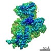 3663MC 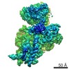 3661C 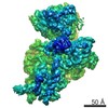 3662C  5no2C 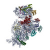 5no3C M: map data used to model this data C: citing same article ( |
|---|---|
| Similar structure data |
-Links
 PDBj
PDBj


- Assembly
| Deposited unit | 
|
|---|---|
| 1 |
|
-Components
-30S ribosomal protein ... , 18 types, 18 molecules CDEFGHIJKLMNOPQRST
| #2: Protein | Mass: 23078.785 Da / Num. of mol.: 1 / Source method: isolated from a natural source / Source: (natural) |
|---|---|
| #3: Protein | Mass: 23383.002 Da / Num. of mol.: 1 / Source method: isolated from a natural source / Source: (natural) |
| #4: Protein | Mass: 16361.878 Da / Num. of mol.: 1 / Source method: isolated from a natural source / Source: (natural) |
| #5: Protein | Mass: 12326.251 Da / Num. of mol.: 1 / Source method: isolated from a natural source / Source: (natural) |
| #6: Protein | Mass: 14543.850 Da / Num. of mol.: 1 / Source method: isolated from a natural source / Source: (natural) |
| #7: Protein | Mass: 14015.361 Da / Num. of mol.: 1 / Source method: isolated from a natural source / Source: (natural) |
| #8: Protein | Mass: 14554.882 Da / Num. of mol.: 1 / Source method: isolated from a natural source / Source: (natural) |
| #9: Protein | Mass: 11325.117 Da / Num. of mol.: 1 / Source method: isolated from a natural source / Source: (natural) |
| #10: Protein | Mass: 12487.200 Da / Num. of mol.: 1 / Source method: isolated from a natural source / Source: (natural) |
| #11: Protein/peptide | Mass: 1641.981 Da / Num. of mol.: 1 / Source method: isolated from a natural source / Source: (natural) |
| #12: Protein | Mass: 12625.753 Da / Num. of mol.: 1 / Source method: isolated from a natural source / Source: (natural) |
| #13: Protein | Mass: 11475.364 Da / Num. of mol.: 1 / Source method: isolated from a natural source / Source: (natural) |
| #14: Protein | Mass: 10159.621 Da / Num. of mol.: 1 / Source method: isolated from a natural source / Source: (natural) |
| #15: Protein | Mass: 9207.572 Da / Num. of mol.: 1 / Source method: isolated from a natural source / Source: (natural) |
| #16: Protein | Mass: 9263.946 Da / Num. of mol.: 1 / Source method: isolated from a natural source / Source: (natural) |
| #17: Protein | Mass: 6466.477 Da / Num. of mol.: 1 / Source method: isolated from a natural source / Source: (natural) |
| #18: Protein | Mass: 9057.626 Da / Num. of mol.: 1 / Source method: isolated from a natural source / Source: (natural) |
| #19: Protein | Mass: 9577.268 Da / Num. of mol.: 1 / Source method: isolated from a natural source / Source: (natural) |
-RNA chain / Protein , 2 types, 2 molecules AZ
| #1: RNA chain | Mass: 497404.969 Da / Num. of mol.: 1 / Source method: isolated from a natural source / Source: (natural) |
|---|---|
| #20: Protein | Mass: 34850.457 Da / Num. of mol.: 1 Source method: isolated from a genetically manipulated source Source: (gene. exp.) Gene: rsgA, engC, yjeQ, b4161, JW4122 / Plasmid: p7XNH3 / Production host: References: UniProt: P39286, Hydrolases; Acting on acid anhydrides; In phosphorus-containing anhydrides |
-Non-polymers , 3 types, 73 molecules 


| #21: Chemical | ChemComp-MG /  Mass: 24.305 Da / Num. of mol.: 71 / Source method: obtained synthetically / Formula: Mg Mass: 24.305 Da / Num. of mol.: 71 / Source method: obtained synthetically / Formula: Mg#22: Chemical | ChemComp-ZN / |  Mass: 65.409 Da / Num. of mol.: 1 / Source method: obtained synthetically / Formula: Zn Mass: 65.409 Da / Num. of mol.: 1 / Source method: obtained synthetically / Formula: Zn#23: Chemical | ChemComp-GNP / |  Mass: 522.196 Da / Num. of mol.: 1 / Source method: obtained synthetically / Formula: C10H17N6O13P3 Mass: 522.196 Da / Num. of mol.: 1 / Source method: obtained synthetically / Formula: C10H17N6O13P3Comment: GppNHp, GMPPNP, energy-carrying molecule analogue*YM |
|---|
-Experimental details
-Experiment
| Experiment | Method: ELECTRON MICROSCOPY |
|---|---|
| EM experiment | Aggregation state: PARTICLE / 3D reconstruction method: single particle reconstruction |
- Sample preparation
| Component | Name: 30S ribosomal subunit bound by RsgA / Type: RIBOSOME / Entity ID: #1-#20 / Source: MULTIPLE SOURCES | |||||||||||||||||||||||||
|---|---|---|---|---|---|---|---|---|---|---|---|---|---|---|---|---|---|---|---|---|---|---|---|---|---|---|
| Molecular weight | Experimental value: NO | |||||||||||||||||||||||||
| Source (natural) | Organism: | |||||||||||||||||||||||||
| Buffer solution | pH: 7.8 | |||||||||||||||||||||||||
| Buffer component |
| |||||||||||||||||||||||||
| Specimen | Embedding applied: NO / Shadowing applied: NO / Staining applied: NO / Vitrification applied: YES | |||||||||||||||||||||||||
| Specimen support | Grid material: COPPER / Grid mesh size: 200 divisions/in. / Grid type: Quantifoil R2/1 | |||||||||||||||||||||||||
| Vitrification | Instrument: FEI VITROBOT MARK II / Cryogen name: ETHANE / Humidity: 95 % / Chamber temperature: 278 K |
- Electron microscopy imaging
| Experimental equipment |  Model: Titan Krios / Image courtesy: FEI Company |
|---|---|
| Microscopy | Model: FEI TITAN KRIOS |
| Electron gun | Electron source:  FIELD EMISSION GUN / Accelerating voltage: 300 kV / Illumination mode: SPOT SCAN FIELD EMISSION GUN / Accelerating voltage: 300 kV / Illumination mode: SPOT SCAN |
| Electron lens | Mode: BRIGHT FIELD / Nominal magnification: 59000 X / Calibrated magnification: 101000 X / Nominal defocus max: 3000 nm / Nominal defocus min: 1000 nm / Cs: 2.7 mm |
| Specimen holder | Cryogen: NITROGEN |
| Image recording | Average exposure time: 1.5 sec. / Electron dose: 2.3 e/Å2 / Detector mode: INTEGRATING / Film or detector model: FEI FALCON II (4k x 4k) / Num. of real images: 3408 |
| Image scans | Width: 2048 / Height: 2048 / Movie frames/image: 13 |
- Processing
| Software | Name: PHENIX / Classification: refinement | |||||||||||||||||||||||||||||||||||||||||||||||||||||||
|---|---|---|---|---|---|---|---|---|---|---|---|---|---|---|---|---|---|---|---|---|---|---|---|---|---|---|---|---|---|---|---|---|---|---|---|---|---|---|---|---|---|---|---|---|---|---|---|---|---|---|---|---|---|---|---|---|
| EM software |
| |||||||||||||||||||||||||||||||||||||||||||||||||||||||
| CTF correction | Type: PHASE FLIPPING AND AMPLITUDE CORRECTION | |||||||||||||||||||||||||||||||||||||||||||||||||||||||
| Particle selection | Num. of particles selected: 878976 | |||||||||||||||||||||||||||||||||||||||||||||||||||||||
| 3D reconstruction | Resolution: 5.16 Å / Resolution method: FSC 0.143 CUT-OFF / Num. of particles: 61908 / Symmetry type: POINT | |||||||||||||||||||||||||||||||||||||||||||||||||||||||
| Atomic model building | Protocol: BACKBONE TRACE | |||||||||||||||||||||||||||||||||||||||||||||||||||||||
| Displacement parameters | Biso max: 486.13 Å2 / Biso mean: 227.5331 Å2 / Biso min: 2.8 Å2 | |||||||||||||||||||||||||||||||||||||||||||||||||||||||
| Refine LS restraints |
|
