- PDB-5lm1: Crystal Structure of HD-PTP phosphatase in complex with UBAP1 -
+
Open data
ID or keywords:
Loading...
-
Basic information
Entry
Database: PDB / ID: 5lm1
Title
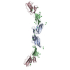
Crystal Structure of HD-PTP phosphatase in complex with UBAP1
Components
Tyrosine-protein phosphatase non-receptor type 23
UBAP-1
Keywords
HYDROLASE / coiled coil
Function / homology
Function and homology information
positive regulation of adherens junction organization / positive regulation of homophilic cell adhesion / ESCRT I complex / positive regulation of Wnt protein secretion / positive regulation of early endosome to late endosome transport / negative regulation of epithelial cell migration / protein transport to vacuole involved in ubiquitin-dependent protein catabolic process via the multivesicular body sorting pathway / early endosome to late endosome transport / membrane fission / ubiquitin-dependent protein catabolic process via the multivesicular body sorting pathway ...positive regulation of adherens junction organization / positive regulation of homophilic cell adhesion / ESCRT I complex / positive regulation of Wnt protein secretion / positive regulation of early endosome to late endosome transport / negative regulation of epithelial cell migration / protein transport to vacuole involved in ubiquitin-dependent protein catabolic process via the multivesicular body sorting pathway / early endosome to late endosome transport / membrane fission / ubiquitin-dependent protein catabolic process via the multivesicular body sorting pathway / multivesicular body assembly / endocytic recycling / Interleukin-37 signaling / cilium assembly / protein-tyrosine-phosphatase / Endosomal Sorting Complex Required For Transport (ESCRT) / Membrane binding and targetting of GAG proteins / protein tyrosine phosphatase activity / ubiquitin binding / HCMV Late Events / Late endosomal microautophagy / Budding and maturation of HIV virion / centriolar satellite / early endosome / endosome / endosome membrane / nuclear body / ciliary basal body / intracellular membrane-bounded organelle / protein kinase binding / extracellular exosome / nucleoplasm / nucleus / plasma membrane / cytoplasm / cytosol Similarity search - Function
Ubiquitin-associated protein 1 / Ubiquitin-associated protein 1, C-terminal / : / Ubiquitin-associated protein 1, UBA2 domain / UBA-like domain / UMA domain / UMA domain profile. / ALIX V-shaped domain / ALIX V-shaped domain binding to HIV / BRO1 domain ...Ubiquitin-associated protein 1 / Ubiquitin-associated protein 1, C-terminal / : / Ubiquitin-associated protein 1, UBA2 domain / UBA-like domain / UMA domain / UMA domain profile. / ALIX V-shaped domain / ALIX V-shaped domain binding to HIV / BRO1 domain / BRO1 domain superfamily / BRO1-like domain / BRO1 domain profile. / BRO1-like domain / Ubiquitin-associated domain / Ubiquitin-associated domain (UBA) profile. / UBA-like superfamily / Protein tyrosine phosphatase, catalytic domain / PTP type protein phosphatase domain profile. / Protein-tyrosine phosphatase / Tyrosine-specific protein phosphatase, PTPase domain / Protein-tyrosine phosphatase, catalytic / Protein tyrosine phosphatase, catalytic domain motif / Tyrosine specific protein phosphatases active site. / Protein-tyrosine phosphatase, active site / Tyrosine specific protein phosphatases domain profile. / Tyrosine-specific protein phosphatases domain / Protein-tyrosine phosphatase-like Similarity search - Domain/homology
In the structure databanks used in Yorodumi, some data are registered as the other names, "COVID-19 virus" and "2019-nCoV". Here are the details of the virus and the list of structure data.
Jan 31, 2019. EMDB accession codes are about to change! (news from PDBe EMDB page)
EMDB accession codes are about to change! (news from PDBe EMDB page)
The allocation of 4 digits for EMDB accession codes will soon come to an end. Whilst these codes will remain in use, new EMDB accession codes will include an additional digit and will expand incrementally as the available range of codes is exhausted. The current 4-digit format prefixed with “EMD-” (i.e. EMD-XXXX) will advance to a 5-digit format (i.e. EMD-XXXXX), and so on. It is currently estimated that the 4-digit codes will be depleted around Spring 2019, at which point the 5-digit format will come into force.
The EM Navigator/Yorodumi systems omit the EMD- prefix.
Related info.:Q: What is EMD? / ID/Accession-code notation in Yorodumi/EM Navigator
Yorodumi is a browser for structure data from EMDB, PDB, SASBDB, etc.
This page is also the successor to EM Navigator detail page, and also detail information page/front-end page for Omokage search.
The word "yorodu" (or yorozu) is an old Japanese word meaning "ten thousand". "mi" (miru) is to see.
Related info.:EMDB / PDB / SASBDB / Comparison of 3 databanks / Yorodumi Search / Aug 31, 2016. New EM Navigator & Yorodumi / Yorodumi Papers / Jmol/JSmol / Function and homology information / Changes in new EM Navigator and Yorodumi
 Movie
Movie Controller


 Open data
 Basic information
 Components
 Keywords
 Function and homology information
 Homo sapiens (human)
 X-RAY DIFFRACTION /
  Authors
 Citation
 Structure visualization
 Downloads & links
 Other downloads

 PDBj
PDBj


 Assembly
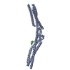

 Mass: 18.015 Da / Num. of mol.: 31 / Source method: isolated from a natural source / Formula: H2O
 Sample preparation
 / Beamline: I02 / Wavelength: 0.979 Å
 Processing