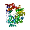
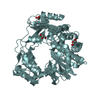
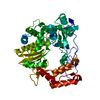
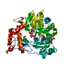
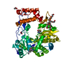
Entry Database : PDB / ID : 4y2cTitle M300V 3D polymerase mutant of EMCV Genome polyprotein Keywords / Function / homology Function Domain/homology Component
/ / / / / / / / / / / / / / / / / / / / / / / / / / / / / / / / / / / / / / / / / / / / / / / / / / / / / / / / / / / / / / / / / / / / / / / / / / / / / / Biological species Method / / / Resolution : 2.2 Å Authors Verdaguer, N. / Ferrer-Orta, C. / Vives-Adrian, L. Journal : Plos Pathog. / Year : 2015Title : The RNA Template Channel of the RNA-Dependent RNA Polymerase as a Target for Development of Antiviral Therapy of Multiple Genera within a Virus Family.Authors: van der Linden, L. / Vives-Adrian, L. / Selisko, B. / Ferrer-Orta, C. / Liu, X. / Lanke, K. / Ulferts, R. / De Palma, A.M. / Tanchis, F. / Goris, N. / Lefebvre, D. / De Clercq, K. / Leyssen, ... Authors : van der Linden, L. / Vives-Adrian, L. / Selisko, B. / Ferrer-Orta, C. / Liu, X. / Lanke, K. / Ulferts, R. / De Palma, A.M. / Tanchis, F. / Goris, N. / Lefebvre, D. / De Clercq, K. / Leyssen, P. / Lacroix, C. / Purstinger, G. / Coutard, B. / Canard, B. / Boehr, D.D. / Arnold, J.J. / Cameron, C.E. / Verdaguer, N. / Neyts, J. / van Kuppeveld, F.J. History Deposition Feb 9, 2015 Deposition site / Processing site Revision 1.0 Apr 1, 2015 Provider / Type Revision 1.1 Jan 10, 2024 Group / Database references / Refinement descriptionCategory chem_comp_atom / chem_comp_bond ... chem_comp_atom / chem_comp_bond / database_2 / pdbx_initial_refinement_model Item / _database_2.pdbx_database_accession
Show all Show less
 Movie
Movie Controller


 Open data
 Basic information
 Components
 Keywords
 Function and homology information
 Mengo encephalomyocarditis virus
 X-RAY DIFFRACTION /
  Authors
 Citation
 Structure visualization
 Downloads & links
 Other downloads
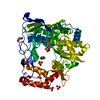


 PDBj
PDBj

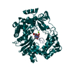
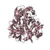
 Assembly


 Mass: 92.094 Da / Num. of mol.: 4 / Source method: obtained synthetically / Formula: C3H8O3
 Mass: 18.015 Da / Num. of mol.: 176 / Source method: isolated from a natural source / Formula: H2O
 Sample preparation
 / Beamline: XALOC / Wavelength: 0.979 Å
 Processing