 Movie
Movie Controller

[English] 日本語
 Yorodumi
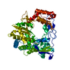
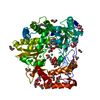
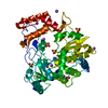
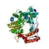
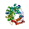
- PDB-4y34: Crystal Structure of Coxsackievirus B3 3D polymerase in complex w... -
+ Open data

- Basic information
| Entry | Database: PDB / ID: 4y34 | ||||||
|---|---|---|---|---|---|---|---|
| Title | Crystal Structure of Coxsackievirus B3 3D polymerase in complex with GPC-N143 | ||||||
 Components Components | 3D polymerase | ||||||
 Keywords Keywords | TRANSFERASE / Polymerase / Picornavirus / Coxsackievirus / GPC-N143 / inhibitor / VIRAL PROTEIN | ||||||
| Function / homology |  Function and homology information Function and homology informationsymbiont-mediated suppression of host cytoplasmic pattern recognition receptor signaling pathway via inhibition of RIG-I activity / picornain 2A / symbiont-mediated suppression of host mRNA export from nucleus / symbiont genome entry into host cell via pore formation in plasma membrane / picornain 3C / T=pseudo3 icosahedral viral capsid / host cell cytoplasmic vesicle membrane / ribonucleoside triphosphate phosphatase activity / nucleoside-triphosphate phosphatase / channel activity ...symbiont-mediated suppression of host cytoplasmic pattern recognition receptor signaling pathway via inhibition of RIG-I activity / picornain 2A / symbiont-mediated suppression of host mRNA export from nucleus / symbiont genome entry into host cell via pore formation in plasma membrane / picornain 3C / T=pseudo3 icosahedral viral capsid / host cell cytoplasmic vesicle membrane / ribonucleoside triphosphate phosphatase activity / nucleoside-triphosphate phosphatase / channel activity / monoatomic ion transmembrane transport / DNA replication / RNA helicase activity / endocytosis involved in viral entry into host cell / symbiont-mediated activation of host autophagy / RNA-directed RNA polymerase / cysteine-type endopeptidase activity / viral RNA genome replication / RNA-directed RNA polymerase activity / DNA-templated transcription / virion attachment to host cell / host cell nucleus / structural molecule activity / proteolysis / RNA binding / zinc ion binding / ATP binding Similarity search - Function | ||||||
| Biological species |   Coxsackievirus B3 Coxsackievirus B3synthetic construct (others) | ||||||
| Method |  X-RAY DIFFRACTION / SYNCHROTRON / MOLECULAR REPLACEMENT / Resolution: 2.7 Å X-RAY DIFFRACTION / SYNCHROTRON / MOLECULAR REPLACEMENT / Resolution: 2.7 Å | ||||||
 Authors Authors | Vives-Adrian, L. / Ferrer-Orta, C. / Cerdaguer, N. | ||||||
 Citation Citation | Journal: Plos Pathog. / Year: 2015 Title: The RNA Template Channel of the RNA-Dependent RNA Polymerase as a Target for Development of Antiviral Therapy of Multiple Genera within a Virus Family. Authors: van der Linden, L. / Vives-Adrian, L. / Selisko, B. / Ferrer-Orta, C. / Liu, X. / Lanke, K. / Ulferts, R. / De Palma, A.M. / Tanchis, F. / Goris, N. / Lefebvre, D. / De Clercq, K. / Leyssen, ...Authors: van der Linden, L. / Vives-Adrian, L. / Selisko, B. / Ferrer-Orta, C. / Liu, X. / Lanke, K. / Ulferts, R. / De Palma, A.M. / Tanchis, F. / Goris, N. / Lefebvre, D. / De Clercq, K. / Leyssen, P. / Lacroix, C. / Purstinger, G. / Coutard, B. / Canard, B. / Boehr, D.D. / Arnold, J.J. / Cameron, C.E. / Verdaguer, N. / Neyts, J. / van Kuppeveld, F.J. | ||||||
| History |
|
- Structure visualization
| Structure viewer | Molecule: MolmilJmol/JSmol |
|---|
- Downloads & links
-Download
| PDBx/mmCIF format | 4y34.cif.gz | 108.4 KB | Display | PDBx/mmCIF format |
|---|---|---|---|---|
| PDB format | pdb4y34.ent.gz | 83.7 KB | Display | PDB format |
| PDBx/mmJSON format | 4y34.json.gz | Tree view | PDBx/mmJSON format | |
| Others |  Other downloads Other downloads |
-Validation report
| Arichive directory | https://data.pdbj.org/pub/pdb/validation_reports/y3/4y34ftp://data.pdbj.org/pub/pdb/validation_reports/y3/4y34 | HTTPS FTP |
|---|
-Related structure data
| Related structure data | 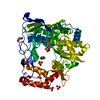 4y2aC  4y2cC  4y3cC  3ddkS S: Starting model for refinement C: citing same article ( |
|---|---|
| Similar structure data |


-Links
 PDBj
PDBj

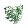
- Assembly
| Deposited unit | 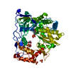
| ||||||||
|---|---|---|---|---|---|---|---|---|---|
| 1 |
| ||||||||
| Unit cell |
|
-Components
| #1: Protein | Mass: 53167.773 Da / Num. of mol.: 1 Source method: isolated from a genetically manipulated source Source: (gene. exp.) Coxsackievirus B3 / Production host:  | ||||
|---|---|---|---|---|---|
| #2: Chemical | ChemComp-45Z /   Mass: 420.307 Da / Num. of mol.: 1 / Source method: obtained synthetically / Formula: C20H9FN4O6 / Source: (synth.) synthetic construct (others) Mass: 420.307 Da / Num. of mol.: 1 / Source method: obtained synthetically / Formula: C20H9FN4O6 / Source: (synth.) synthetic construct (others) | ||||
| #3: Chemical | ChemComp-SO4 /   Mass: 96.063 Da / Num. of mol.: 5 / Source method: obtained synthetically / Formula: SO4 Mass: 96.063 Da / Num. of mol.: 5 / Source method: obtained synthetically / Formula: SO4#4: Chemical | ChemComp-GOL /   Mass: 92.094 Da / Num. of mol.: 4 / Source method: obtained synthetically / Formula: C3H8O3 Mass: 92.094 Da / Num. of mol.: 4 / Source method: obtained synthetically / Formula: C3H8O3#5: Water | ChemComp-HOH / |  Mass: 18.015 Da / Num. of mol.: 56 / Source method: isolated from a natural source / Formula: H2O Mass: 18.015 Da / Num. of mol.: 56 / Source method: isolated from a natural source / Formula: H2O |
-Experimental details
-Experiment
| Experiment | Method: X-RAY DIFFRACTION |
|---|
- Sample preparation
| Crystal | Density Matthews: 3.75 Å3/Da / Density % sol: 67.17 % |
|---|---|
| Crystal grow | Temperature: 293 K / Method: vapor diffusion, sitting drop Details: 50mM Tris pH 7.5, 24.5% (w/v) Glycerol, 1,29M Ammonium Sulfate |
-Data collection
| Diffraction | Mean temperature: 100 K |
|---|---|
| Diffraction source | Source: SYNCHROTRON / Site: ESRF  / Beamline: ID29 / Wavelength: 0.977 Å / Beamline: ID29 / Wavelength: 0.977 Å |
| Detector | Type: PSI PILATUS 6M / Detector: PIXEL / Date: Jul 8, 2011 |
| Radiation | Protocol: SINGLE WAVELENGTH / Monochromatic (M) / Laue (L): M / Scattering type: x-ray |
| Radiation wavelength | Wavelength: 0.977 Å / Relative weight: 1 |
| Reflection | Resolution: 2.7→72.2 Å / Num. obs: 21638 / % possible obs: 96.8 % / Redundancy: 3.6 % / Rmerge(I) obs: 0.072 / Rsym value: 0.07 / Net I/σ(I): 9.2 |
| Reflection shell | Resolution: 2.7→2.85 Å / Redundancy: 3.3 % / Rmerge(I) obs: 0.471 / Rsym value: 0.4 / % possible all: 95.8 |
- Processing
| Software |
| ||||||||||||||||||||||||||||||||||||||||||||||||||||||||||||||||||||||||||||||||||||||||||||||||||||||||||||||||||||||||||||||||||||||||||||||||||||||||||||||||||||||||||||||||||||||
|---|---|---|---|---|---|---|---|---|---|---|---|---|---|---|---|---|---|---|---|---|---|---|---|---|---|---|---|---|---|---|---|---|---|---|---|---|---|---|---|---|---|---|---|---|---|---|---|---|---|---|---|---|---|---|---|---|---|---|---|---|---|---|---|---|---|---|---|---|---|---|---|---|---|---|---|---|---|---|---|---|---|---|---|---|---|---|---|---|---|---|---|---|---|---|---|---|---|---|---|---|---|---|---|---|---|---|---|---|---|---|---|---|---|---|---|---|---|---|---|---|---|---|---|---|---|---|---|---|---|---|---|---|---|---|---|---|---|---|---|---|---|---|---|---|---|---|---|---|---|---|---|---|---|---|---|---|---|---|---|---|---|---|---|---|---|---|---|---|---|---|---|---|---|---|---|---|---|---|---|---|---|---|---|
| Refinement | Method to determine structure: MOLECULAR REPLACEMENT Starting model: 3DDK Resolution: 2.7→72.2 Å / Cor.coef. Fo:Fc: 0.933 / Cor.coef. Fo:Fc free: 0.91 / SU B: 9.984 / SU ML: 0.205 / Cross valid method: THROUGHOUT / ESU R: 0.455 / ESU R Free: 0.283 / Stereochemistry target values: MAXIMUM LIKELIHOOD / Details: HYDROGENS HAVE BEEN ADDED IN THE RIDING POSITIONS
| ||||||||||||||||||||||||||||||||||||||||||||||||||||||||||||||||||||||||||||||||||||||||||||||||||||||||||||||||||||||||||||||||||||||||||||||||||||||||||||||||||||||||||||||||||||||
| Solvent computation | Ion probe radii: 0.8 Å / Shrinkage radii: 0.8 Å / VDW probe radii: 1.4 Å / Solvent model: MASK | ||||||||||||||||||||||||||||||||||||||||||||||||||||||||||||||||||||||||||||||||||||||||||||||||||||||||||||||||||||||||||||||||||||||||||||||||||||||||||||||||||||||||||||||||||||||
| Displacement parameters | Biso mean: 49.647 Å2
| ||||||||||||||||||||||||||||||||||||||||||||||||||||||||||||||||||||||||||||||||||||||||||||||||||||||||||||||||||||||||||||||||||||||||||||||||||||||||||||||||||||||||||||||||||||||
| Refinement step | Cycle: 1 / Resolution: 2.7→72.2 Å
| ||||||||||||||||||||||||||||||||||||||||||||||||||||||||||||||||||||||||||||||||||||||||||||||||||||||||||||||||||||||||||||||||||||||||||||||||||||||||||||||||||||||||||||||||||||||
| Refine LS restraints |
|