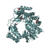
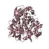
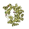
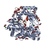
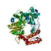
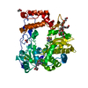
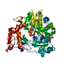
[English] 日本語
Yorodumi
- PDB-4nyz: The EMCV 3Dpol structure with altered motif A conformation at 2.1... -

+
Open data


ID or keywords:

Loading...

-
Basic information

Entry
Database: PDB / ID: 4nyz
TitleThe EMCV 3Dpol structure with altered motif A conformation at 2.15A resolution
ComponentsGenome polyprotein
KeywordsTRANSFERASE / Encephalomyocarditis virus / Close right hand / RNa dependent RNA polymerase
Function / homology
Function and homology information


positive stranded viral RNA replication / host cell nucleolus / symbiont-mediated suppression of host cytoplasmic pattern recognition receptor signaling pathway via inhibition of RIG-I activity / picornain 3C / T=pseudo3 icosahedral viral capsid / host cell cytoplasmic vesicle membrane / : / protein complex oligomerization / monoatomic ion channel activity / RNA helicase activity ...positive stranded viral RNA replication / host cell nucleolus / symbiont-mediated suppression of host cytoplasmic pattern recognition receptor signaling pathway via inhibition of RIG-I activity / picornain 3C / T=pseudo3 icosahedral viral capsid / host cell cytoplasmic vesicle membrane / : / protein complex oligomerization / monoatomic ion channel activity / RNA helicase activity / RNA helicase / induction by virus of host autophagy / symbiont entry into host cell / RNA-directed RNA polymerase / symbiont-mediated suppression of host gene expression / cysteine-type endopeptidase activity / RNA-dependent RNA polymerase activity / DNA-templated transcription / structural molecule activity / virion attachment to host cell / ATP hydrolysis activity / proteolysis / RNA binding / ATP binding / membrane / metal ion binding
Similarity search - Function
Leader peptide, picornavirus / Viral leader polypeptide zinc finger / Virion protein N terminal domain / Capsid protein VP4, Picornavirus / Viral protein VP4 subunit / Mitochondrial Import Receptor Subunit Tom20; Chain A - #20 / Capsid protein VP4 superfamily, Picornavirus / Mitochondrial Import Receptor Subunit Tom20; Chain A / Helicase/polymerase/peptidase polyprotein, Calicivirus-type / Reverse transcriptase/Diguanylate cyclase domain ...Leader peptide, picornavirus / Viral leader polypeptide zinc finger / Virion protein N terminal domain / Capsid protein VP4, Picornavirus / Viral protein VP4 subunit / Mitochondrial Import Receptor Subunit Tom20; Chain A - #20 / Capsid protein VP4 superfamily, Picornavirus / Mitochondrial Import Receptor Subunit Tom20; Chain A / Helicase/polymerase/peptidase polyprotein, Calicivirus-type / Reverse transcriptase/Diguanylate cyclase domain / Picornavirales 3C/3C-like protease domain / Picornavirales 3C/3C-like protease domain profile. / Peptidase C3A/C3B, picornaviral / 3C cysteine protease (picornain 3C) / Picornavirus capsid / picornavirus capsid protein / Helicase, superfamily 3, single-stranded RNA virus / Superfamily 3 helicase of positive ssRNA viruses domain profile. / Helicase, superfamily 3, single-stranded DNA/RNA virus / RNA helicase / Picornavirus/Calicivirus coat protein / Viral coat protein subunit / RNA-directed RNA polymerase, C-terminal domain / Viral RNA-dependent RNA polymerase / Reverse transcriptase/Diguanylate cyclase domain / RNA-directed RNA polymerase, catalytic domain / RdRp of positive ssRNA viruses catalytic domain profile. / Alpha-Beta Plaits / Peptidase S1, PA clan, chymotrypsin-like fold / Peptidase S1, PA clan / DNA/RNA polymerase superfamily / Up-down Bundle / 2-Layer Sandwich / Mainly Alpha / Alpha Beta
Similarity search - Domain/homology
GLUTAMINE / Genome polyprotein
Similarity search - Component
Biological speciesMengo virus
MethodX-RAY DIFFRACTION / SYNCHROTRON / MOLECULAR REPLACEMENT / Resolution: 2.15 Å
AuthorsVives-adrian, L. / Lujan, C. / Ferrer-orta, C. / Verdaguer, N.
CitationJournal: J.Virol. / Year: 2014
Title: The crystal structure of a cardiovirus RNA-dependent RNA polymerase reveals an unusual conformation of the polymerase active site
Authors: Vives-Adrian, L. / Lujan, C. / Oliva, B. / van der Linden, L. / Selisko, B. / Coutard, B. / Canard, B. / van Kuppeveld, F.J. / Ferrer-Orta, C. / Verdaguer, N.
History
DepositionDec 11, 2013Deposition site: RCSB / Processing site: PDBJ
Revision 1.0Mar 19, 2014Provider: repository / Type: Initial release
Revision 1.1Dec 10, 2014Group: Database references
Revision 1.2Nov 8, 2023Group: Data collection / Database references ...Data collection / Database references / Derived calculations / Refinement description
Category: chem_comp_atom / chem_comp_bond ...chem_comp_atom / chem_comp_bond / database_2 / pdbx_initial_refinement_model / pdbx_struct_conn_angle / struct_conn / struct_site
Item: _database_2.pdbx_DOI / _database_2.pdbx_database_accession ..._database_2.pdbx_DOI / _database_2.pdbx_database_accession / _pdbx_struct_conn_angle.ptnr1_auth_comp_id / _pdbx_struct_conn_angle.ptnr1_auth_seq_id / _pdbx_struct_conn_angle.ptnr1_label_atom_id / _pdbx_struct_conn_angle.ptnr1_label_comp_id / _pdbx_struct_conn_angle.ptnr1_label_seq_id / _pdbx_struct_conn_angle.ptnr3_auth_comp_id / _pdbx_struct_conn_angle.ptnr3_auth_seq_id / _pdbx_struct_conn_angle.ptnr3_label_atom_id / _pdbx_struct_conn_angle.ptnr3_label_comp_id / _pdbx_struct_conn_angle.ptnr3_label_seq_id / _struct_conn.pdbx_dist_value / _struct_conn.ptnr1_auth_comp_id / _struct_conn.ptnr1_auth_seq_id / _struct_conn.ptnr1_label_atom_id / _struct_conn.ptnr1_label_comp_id / _struct_conn.ptnr1_label_seq_id / _struct_site.pdbx_auth_asym_id / _struct_site.pdbx_auth_comp_id / _struct_site.pdbx_auth_seq_id

-
Structure visualization

Structure viewerMolecule:
MolmilJmol/JSmol

Downloads & links

-
Assembly

Deposited unit
A: Genome polyprotein
hetero molecules


Theoretical massNumber of molelcules
Total (without water)52,4754
Polymers52,2121
Non-polymers2633
Water1,982110
1


  • Idetical with deposited unit
  • defined by author&software
TypeNameSymmetry operationNumber
identity operation1_555x,y,z1
MethodPISA
Unit cell
Length a, b, c (Å)122.553, 122.553, 198.797
Angle α, β, γ (deg.)90.00, 90.00, 90.00
Int Tables number98
Space group name H-MI4122
Components on special symmetry positions
IDModelComponents
11A-612-

HOH

21A-658-

HOH

-
Components

#1: Protein Genome polyprotein / Leader protein / Protein VP0 / VP4-VP2 / Protein VP4 / P1A / Rho / Virion protein 4 / Protein VP2 / ...Leader protein / Protein VP0 / VP4-VP2 / Protein VP4 / P1A / Rho / Virion protein 4 / Protein VP2 / Beta / P1B / Virion protein 2 / Protein VP3 / Gamma / P1C / Virion protein 3 / Protein VP1 / Alpha / P1D / Virion protein 1 / Protein 2A / P2A / G / Protein 2B / I / P2B / Protein 2C / C / P2C / Protein 3A / P3A / Protein 3B / P3B / H / VPg / Protease 3C / P3C / Picornain 3C / p22 / RNA-directed RNA polymerase 3D-POL / E / P3D-POL


Mass: 52212.441 Da / Num. of mol.: 1 / Fragment: UNP residues 1834-2293
Source method: isolated from a genetically manipulated source
Source: (gene. exp.) Mengo virus / Plasmid: pETG20A / Production host: Escherichia coli (E. coli) / Strain (production host): Rosetta (DE3) pLyS
References: UniProt: P12296, nucleoside-triphosphate phosphatase, picornain 3C, RNA-directed RNA polymerase
#2: Chemical ChemComp-GLN / GLUTAMINE / Glutamine


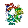
Type: L-peptide linking / Mass: 146.144 Da / Num. of mol.: 1 / Source method: obtained synthetically / Formula: C5H10N2O3
#3: Chemical ChemComp-MG / MAGNESIUM ION


Mass: 24.305 Da / Num. of mol.: 1 / Source method: obtained synthetically / Formula: Mg
#4: Chemical ChemComp-GOL / GLYCEROL / GLYCERIN / PROPANE-1,2,3-TRIOL / Glycerol


Mass: 92.094 Da / Num. of mol.: 1 / Source method: obtained synthetically / Formula: C3H8O3
#5: Water ChemComp-HOH / water / Water


Mass: 18.015 Da / Num. of mol.: 110 / Source method: isolated from a natural source / Formula: H2O

-
Experimental details

-
Experiment

ExperimentMethod: X-RAY DIFFRACTION / Number of used crystals: 1

-
Sample preparation

CrystalDensity Matthews: 3.57 Å3/Da / Density % sol: 65.59 %
Crystal growTemperature: 277 K / Method: vapor diffusion, sitting drop / pH: 7.5
Details: 10%PEG6000 and 2.0M NaCl, pH 7.5, VAPOR DIFFUSION, SITTING DROP, temperature 277K

-
Data collection

DiffractionMean temperature: 100 K
Diffraction sourceSource: SYNCHROTRON / Site: ALBA / Beamline: XALOC / Wavelength: 0.979 Å
DetectorType: PSI PILATUS 6M / Detector: PIXEL / Date: Feb 2, 2013
RadiationMonochromator: Channel-cut Si(111) / Protocol: SINGLE WAVELENGTH / Monochromatic (M) / Laue (L): M / Scattering type: x-ray
Radiation wavelengthWavelength: 0.979 Å / Relative weight: 1
ReflectionResolution: 2.15→104.3 Å / Num. all: 54658 / Num. obs: 40499 / % possible obs: 98.4 % / Observed criterion σ(F): 2 / Observed criterion σ(I): 2 / Redundancy: 5 % / Rmerge(I) obs: 0.11 / Rsym value: 0.099 / Net I/σ(I): 6.4
Reflection shellResolution: 2.15→2.27 Å / Redundancy: 5 % / Rmerge(I) obs: 0.86 / Mean I/σ(I) obs: 18.7 / Rsym value: 0.77 / % possible all: 98.3

-
Processing

Software
NameVersionClassification
PHASERphasing
REFMAC5.8.0049refinement
XDSdata reduction
SCALAdata scaling
RefinementMethod to determine structure: MOLECULAR REPLACEMENT
Starting model: 1U09(THE HOMOLOGY MODEL HAS BEEN USED.)
Resolution: 2.15→104.3 Å / Cor.coef. Fo:Fc: 0.934 / Cor.coef. Fo:Fc free: 0.92 / SU B: 11.575 / SU ML: 0.146 / Cross valid method: THROUGHOUT / ESU R: 0.206 / ESU R Free: 0.175 / Stereochemistry target values: MAXIMUM LIKELIHOOD / Details: HYDROGENS HAVE BEEN ADDED IN THE RIDING POSITIONS
RfactorNum. reflection% reflectionSelection details
Rfree0.2522 2036 5 %RANDOM
Rwork0.22948 ---
all0.23061 54658 --
obs0.23061 38457 97.73 %-
Solvent computationIon probe radii: 0.8 Å / Shrinkage radii: 0.8 Å / VDW probe radii: 1.2 Å / Solvent model: MASK
Displacement parametersBiso mean: 40.315 Å2
Baniso -1Baniso -2Baniso -3
1-0.96 Å2-0 Å20 Å2
2--0.96 Å2-0 Å2
3----1.93 Å2
Refinement stepCycle: LAST / Resolution: 2.15→104.3 Å
ProteinNucleic acidLigandSolventTotal
Num. atoms3680 0 17 110 3807
Refine LS restraints
Refine-IDTypeDev idealDev ideal targetNumber
X-RAY DIFFRACTIONr_bond_refined_d0.0040.0193806
X-RAY DIFFRACTIONr_bond_other_d0.0010.023623
X-RAY DIFFRACTIONr_angle_refined_deg0.8771.9715158
X-RAY DIFFRACTIONr_angle_other_deg0.738337
X-RAY DIFFRACTIONr_dihedral_angle_1_deg4.7275463
X-RAY DIFFRACTIONr_dihedral_angle_2_deg30.17223.333180
X-RAY DIFFRACTIONr_dihedral_angle_3_deg14.2415646
X-RAY DIFFRACTIONr_dihedral_angle_4_deg14.7871531
X-RAY DIFFRACTIONr_chiral_restr0.0520.2567
X-RAY DIFFRACTIONr_gen_planes_refined0.0030.0214286
X-RAY DIFFRACTIONr_gen_planes_other0.0010.02891
X-RAY DIFFRACTIONr_nbd_refined
X-RAY DIFFRACTIONr_nbd_other
X-RAY DIFFRACTIONr_nbtor_refined
X-RAY DIFFRACTIONr_nbtor_other
X-RAY DIFFRACTIONr_xyhbond_nbd_refined
X-RAY DIFFRACTIONr_xyhbond_nbd_other
X-RAY DIFFRACTIONr_metal_ion_refined
X-RAY DIFFRACTIONr_metal_ion_other
X-RAY DIFFRACTIONr_symmetry_vdw_refined
X-RAY DIFFRACTIONr_symmetry_vdw_other
X-RAY DIFFRACTIONr_symmetry_hbond_refined
X-RAY DIFFRACTIONr_symmetry_hbond_other
X-RAY DIFFRACTIONr_symmetry_metal_ion_refined
X-RAY DIFFRACTIONr_symmetry_metal_ion_other
X-RAY DIFFRACTIONr_mcbond_it0.5282.4781852
X-RAY DIFFRACTIONr_mcbond_other0.5282.4771851
X-RAY DIFFRACTIONr_mcangle_it0.9763.7132315
X-RAY DIFFRACTIONr_mcangle_other0.9763.7142316
X-RAY DIFFRACTIONr_scbond_it0.4132.4941953
X-RAY DIFFRACTIONr_scbond_other0.4142.4941953
X-RAY DIFFRACTIONr_scangle_it
X-RAY DIFFRACTIONr_scangle_other0.753.7212844
X-RAY DIFFRACTIONr_long_range_B_refined2.87119.5154274
X-RAY DIFFRACTIONr_long_range_B_other2.87119.5194275
X-RAY DIFFRACTIONr_rigid_bond_restr
X-RAY DIFFRACTIONr_sphericity_free
X-RAY DIFFRACTIONr_sphericity_bonded
LS refinement shellResolution: 2.15→2.206 Å / Total num. of bins used: 20
RfactorNum. reflection% reflection
Rfree0.307 146 -
Rwork0.307 2816 -
obs--98.31 %
Refinement TLS params.Method: refined / Origin x: -7.6644 Å / Origin y: -29.2899 Å / Origin z: -23.9081 Å
111213212223313233
T0.0629 Å2-0.0018 Å20.0182 Å2-0.0695 Å20.0033 Å2--0.035 Å2
L0.6692 °2-0.2524 °20.3984 °2-1.3987 °2-0.0213 °2--2.0261 °2
S-0.0219 Å °0.0185 Å °0.1301 Å °-0.1688 Å °-0.0111 Å °-0.0899 Å °-0.0865 Å °0.3273 Å °0.033 Å °

+
About Yorodumi

-
News

-
Feb 9, 2022. New format data for meta-information of EMDB entries

New format data for meta-information of EMDB entries

  • Version 3 of the EMDB header file is now the official format.
  • The previous official version 1.9 will be removed from the archive.

Related info.:EMDB header

External links:wwPDB to switch to version 3 of the EMDB data model

-
Aug 12, 2020. Covid-19 info

Covid-19 info

URL: https://pdbj.org/emnavi/covid19.php

New page: Covid-19 featured information page in EM Navigator.

Related info.:Covid-19 info / Mar 5, 2020. Novel coronavirus structure data

+
Mar 5, 2020. Novel coronavirus structure data

Novel coronavirus structure data

Related info.:Yorodumi Speices / Aug 12, 2020. Covid-19 info

External links:COVID-19 featured content - PDBj / Molecule of the Month (242):Coronavirus Proteases

+
Jan 31, 2019. EMDB accession codes are about to change! (news from PDBe EMDB page)

EMDB accession codes are about to change! (news from PDBe EMDB page)

  • The allocation of 4 digits for EMDB accession codes will soon come to an end. Whilst these codes will remain in use, new EMDB accession codes will include an additional digit and will expand incrementally as the available range of codes is exhausted. The current 4-digit format prefixed with “EMD-” (i.e. EMD-XXXX) will advance to a 5-digit format (i.e. EMD-XXXXX), and so on. It is currently estimated that the 4-digit codes will be depleted around Spring 2019, at which point the 5-digit format will come into force.
  • The EM Navigator/Yorodumi systems omit the EMD- prefix.

Related info.:Q: What is EMD? / ID/Accession-code notation in Yorodumi/EM Navigator

External links:EMDB Accession Codes are Changing Soon! / Contact to PDBj

+
Jul 12, 2017. Major update of PDB

Major update of PDB

  • wwPDB released updated PDB data conforming to the new PDBx/mmCIF dictionary.
  • This is a major update changing the version number from 4 to 5, and with Remediation, in which all the entries are updated.
  • In this update, many items about electron microscopy experimental information are reorganized (e.g. em_software).
  • Now, EM Navigator and Yorodumi are based on the updated data.

External links:wwPDB Remediation / Enriched Model Files Conforming to OneDep Data Standards Now Available in the PDB FTP Archive

-
Yorodumi

Thousand views of thousand structures

  • Yorodumi is a browser for structure data from EMDB, PDB, SASBDB, etc.
  • This page is also the successor to EM Navigator detail page, and also detail information page/front-end page for Omokage search.
  • The word "yorodu" (or yorozu) is an old Japanese word meaning "ten thousand". "mi" (miru) is to see.

Related info.:EMDB / PDB / SASBDB / Comparison of 3 databanks / Yorodumi Search / Aug 31, 2016. New EM Navigator & Yorodumi / Yorodumi Papers / Jmol/JSmol / Function and homology information / Changes in new EM Navigator and Yorodumi

Read more