 Movie
Movie Controller

[English] 日本語
 Yorodumi
- PDB-3cdw: Crystal structure of coxsackievirus B3 RNA-dependent RNA polymera... -
+ Open data

- Basic information
| Entry | Database: PDB / ID: 3cdw | ||||||
|---|---|---|---|---|---|---|---|
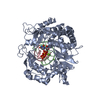
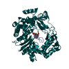
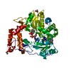
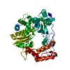
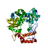
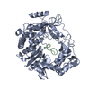
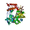
| Title | Crystal structure of coxsackievirus B3 RNA-dependent RNA polymerase (3Dpol) in complex with protein primer VPg and a pyrophosphate | ||||||
 Components Components |
| ||||||
 Keywords Keywords | Transferase/VIRAL PROTEIN / coxsackievirus / RNA-dependent RNA polymerase / protein primer / VPg / VIZIER Viral Enzymes Involved in Replication / Transferase-VIRAL PROTEIN COMPLEX | ||||||
| Function / homology |  Function and homology information Function and homology informationsymbiont-mediated perturbation of host transcription / symbiont-mediated suppression of host cytoplasmic pattern recognition receptor signaling pathway via inhibition of RIG-I activity / symbiont-mediated suppression of host cytoplasmic pattern recognition receptor signaling pathway via inhibition of MDA-5 activity / symbiont-mediated suppression of host cytoplasmic pattern recognition receptor signaling pathway via inhibition of MAVS activity / picornain 2A / symbiont-mediated suppression of host mRNA export from nucleus / symbiont genome entry into host cell via pore formation in plasma membrane / picornain 3C / T=pseudo3 icosahedral viral capsid / host cell cytoplasmic vesicle membrane ...symbiont-mediated perturbation of host transcription / symbiont-mediated suppression of host cytoplasmic pattern recognition receptor signaling pathway via inhibition of RIG-I activity / symbiont-mediated suppression of host cytoplasmic pattern recognition receptor signaling pathway via inhibition of MDA-5 activity / symbiont-mediated suppression of host cytoplasmic pattern recognition receptor signaling pathway via inhibition of MAVS activity / picornain 2A / symbiont-mediated suppression of host mRNA export from nucleus / symbiont genome entry into host cell via pore formation in plasma membrane / picornain 3C / T=pseudo3 icosahedral viral capsid / host cell cytoplasmic vesicle membrane / ribonucleoside triphosphate phosphatase activity / nucleoside-triphosphate phosphatase / channel activity / monoatomic ion transmembrane transport / symbiont-mediated suppression of host NF-kappaB cascade / DNA replication / RNA helicase activity / endocytosis involved in viral entry into host cell / symbiont-mediated activation of host autophagy / RNA-directed RNA polymerase / cysteine-type endopeptidase activity / viral RNA genome replication / RNA-directed RNA polymerase activity / virion attachment to host cell / DNA-templated transcription / host cell nucleus / structural molecule activity / proteolysis / RNA binding / zinc ion binding / ATP binding Similarity search - Function | ||||||
| Biological species |   Coxsackievirus B3 Coxsackievirus B3 | ||||||
| Method |  X-RAY DIFFRACTION / SYNCHROTRON / MOLECULAR REPLACEMENT / Resolution: 2.5 Å X-RAY DIFFRACTION / SYNCHROTRON / MOLECULAR REPLACEMENT / Resolution: 2.5 Å | ||||||
 Authors Authors | Gruez, A. / Selisko, B. / Roberts, M. / Bricogne, G. / Bussetta, C. / Canard, B. | ||||||
 Citation Citation | Journal: J.Virol. / Year: 2008 Title: The crystal structure of coxsackievirus B3 RNA-dependent RNA polymerase in complex with its protein primer VPg confirms the existence of a second VPg binding site on Picornaviridae polymerases Authors: Gruez, A. / Selisko, B. / Roberts, M. / Bricogne, G. / Bussetta, C. / Jabafi, I. / Coutard, B. / De Palma, A.M. / Neyts, J. / Canard, B. | ||||||
| History |
|
- Structure visualization
| Structure viewer | Molecule: MolmilJmol/JSmol |
|---|
- Downloads & links
-Download
| PDBx/mmCIF format | 3cdw.cif.gz | 123.5 KB | Display | PDBx/mmCIF format |
|---|---|---|---|---|
| PDB format | pdb3cdw.ent.gz | 95.7 KB | Display | PDB format |
| PDBx/mmJSON format | 3cdw.json.gz | Tree view | PDBx/mmJSON format | |
| Others |  Other downloads Other downloads |
-Validation report
| Arichive directory | https://data.pdbj.org/pub/pdb/validation_reports/cd/3cdwftp://data.pdbj.org/pub/pdb/validation_reports/cd/3cdw | HTTPS FTP |
|---|
-Related structure data
| Related structure data |  3cduSC S: Starting model for refinement C: citing same article ( |
|---|---|
| Similar structure data |


-Links
 PDBj
PDBj


- Assembly
| Deposited unit | 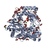
| ||||||||
|---|---|---|---|---|---|---|---|---|---|
| 1 |
| ||||||||
| Unit cell |
|
-Components
-Protein / Protein/peptide , 2 types, 2 molecules AH
| #1: Protein | Mass: 53277.867 Da / Num. of mol.: 1 Source method: isolated from a genetically manipulated source Source: (gene. exp.) Coxsackievirus B3 / Strain: Nancy / Plasmid: pDest14 / Production host:  |
|---|---|
| #2: Protein/peptide | Mass: 2412.787 Da / Num. of mol.: 1 / Source method: obtained synthetically / Details: peptide synthesis / References: UniProt: P03313 |
-Non-polymers , 5 types, 359 molecules 


| #3: Chemical | ChemComp-CL /  Mass: 35.453 Da / Num. of mol.: 7 / Source method: obtained synthetically / Formula: Cl Mass: 35.453 Da / Num. of mol.: 7 / Source method: obtained synthetically / Formula: Cl#4: Chemical | ChemComp-ACT /  Mass: 59.044 Da / Num. of mol.: 6 / Source method: obtained synthetically / Formula: C2H3O2 Mass: 59.044 Da / Num. of mol.: 6 / Source method: obtained synthetically / Formula: C2H3O2#5: Chemical | ChemComp-POP / |  Mass: 175.959 Da / Num. of mol.: 1 / Source method: obtained synthetically / Formula: H2O7P2 Mass: 175.959 Da / Num. of mol.: 1 / Source method: obtained synthetically / Formula: H2O7P2#6: Chemical | ChemComp-GOL /  Mass: 92.094 Da / Num. of mol.: 27 / Source method: obtained synthetically / Formula: C3H8O3 Mass: 92.094 Da / Num. of mol.: 27 / Source method: obtained synthetically / Formula: C3H8O3#7: Water | ChemComp-HOH / | Mass: 18.015 Da / Num. of mol.: 318 / Source method: isolated from a natural source / Formula: H2O |
|---|
-Experimental details
-Experiment
| Experiment | Method: X-RAY DIFFRACTION / Number of used crystals: 2 |
|---|
- Sample preparation
| Crystal | Density Matthews: 3.53 Å3/Da / Density % sol: 65.17 % |
|---|---|
| Crystal grow | Temperature: 293 K / Method: vapor diffusion, hanging drop / pH: 10 Details: 2M ammonium sulfate, 0.1M CAPS, 0.2M lithium sulfate, pH 10, VAPOR DIFFUSION, HANGING DROP, temperature 293K |
-Data collection
| Diffraction | Mean temperature: 100 K |
|---|---|
| Diffraction source | Source: SYNCHROTRON / Site: ESRF  / Beamline: ID14-3 / Wavelength: 0.933 Å / Beamline: ID14-3 / Wavelength: 0.933 Å |
| Detector | Type: MAR CCD 165 mm / Detector: CCD / Date: Feb 18, 2006 |
| Radiation | Monochromator: Diamond (111) - Ge (220) / Protocol: SINGLE WAVELENGTH / Monochromatic (M) / Laue (L): M / Scattering type: x-ray |
| Radiation wavelength | Wavelength: 0.933 Å / Relative weight: 1 |
| Reflection | Resolution: 2.5→27.45 Å / Num. all: 28820 / Num. obs: 28791 / % possible obs: 99.9 % / Observed criterion σ(F): 2 / Observed criterion σ(I): 2 / Redundancy: 6.45 % / Biso Wilson estimate: 56.65 Å2 / Rsym value: 0.084 / Net I/σ(I): 17 |
| Reflection shell | Resolution: 2.5→2.64 Å / Redundancy: 4 % / Mean I/σ(I) obs: 2.6 / Rsym value: 0.386 / % possible all: 99.7 |
- Processing
| Software |
| ||||||||||||||||||||||||||||||||||||||||
|---|---|---|---|---|---|---|---|---|---|---|---|---|---|---|---|---|---|---|---|---|---|---|---|---|---|---|---|---|---|---|---|---|---|---|---|---|---|---|---|---|---|
| Refinement | Method to determine structure: MOLECULAR REPLACEMENT Starting model: 3CDU Resolution: 2.5→27.45 Å / Cross valid method: THROUGHOUT / σ(F): 0 / Stereochemistry target values: Engh & Huber
| ||||||||||||||||||||||||||||||||||||||||
| Displacement parameters | Biso mean: 41.3 Å2
| ||||||||||||||||||||||||||||||||||||||||
| Refine analyze | Luzzati coordinate error obs: 0.2715 Å | ||||||||||||||||||||||||||||||||||||||||
| Refinement step | Cycle: LAST / Resolution: 2.5→27.45 Å
| ||||||||||||||||||||||||||||||||||||||||
| Refine LS restraints |
| ||||||||||||||||||||||||||||||||||||||||
| LS refinement shell | Resolution: 2.5→2.65 Å / Total num. of bins used: 9
|
