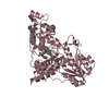
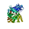
Entry Database : PDB / ID : 4tptTitle Crystal Structure of the Human LIMK2 Kinase Domain In Complex With a Non-ATP Competitive Inhibitor LIM domain kinase 2 Keywords / / / Function / homology Function Domain/homology Component
/ / / / / / / / / / / / / / / / / / / / / / / / / / / / / / / / / / / / / / / / / / / / / / / / / / / / / / / / / / / / / / / / / / Biological species Homo sapiens (human)Method / / / Resolution : 2.6 Å Authors Goodwin, N.C. / Cianchetta, G. / Hamman, B.L. / Burgoon, H.A. / Healy, J. / Mabon, S. / Strobel, E.D. / Wang, S. / Rawlins, D.B. Journal : Acs Med.Chem.Lett. / Year : 2015Title : Discovery of a Type III Inhibitor of LIM Kinase 2 That Binds in a DFG-Out Conformation.Authors : Goodwin, N.C. / Cianchetta, G. / Burgoon, H.A. / Healy, J. / Mabon, R. / Strobel, E.D. / Allen, J. / Wang, S. / Hamman, B.D. / Rawlins, D.B. History Deposition Jun 9, 2014 Deposition site / Processing site Revision 1.0 Oct 22, 2014 Provider / Type Revision 1.1 Jan 28, 2015 Group Revision 2.0 Dec 27, 2023 Group Atomic model / Data collection ... Atomic model / Data collection / Database references / Derived calculations / Other / Refinement description / Source and taxonomy / Structure summary Category atom_site_anisotrop / chem_comp_atom ... atom_site_anisotrop / chem_comp_atom / chem_comp_bond / database_2 / entity_src_gen / pdbx_database_status / pdbx_prerelease_seq / pdbx_struct_assembly / pdbx_struct_assembly_gen / pdbx_struct_assembly_prop / pdbx_struct_oper_list / refine_hist / struct_keywords Item _atom_site_anisotrop.pdbx_PDB_model_num / _atom_site_anisotrop.pdbx_label_asym_id ... _atom_site_anisotrop.pdbx_PDB_model_num / _atom_site_anisotrop.pdbx_label_asym_id / _atom_site_anisotrop.pdbx_label_atom_id / _atom_site_anisotrop.pdbx_label_comp_id / _atom_site_anisotrop.pdbx_label_seq_id / _database_2.pdbx_DOI / _database_2.pdbx_database_accession / _entity_src_gen.pdbx_alt_source_flag / _pdbx_database_status.pdb_format_compatible / _pdbx_struct_assembly.oligomeric_details / _pdbx_struct_assembly_gen.asym_id_list / _pdbx_struct_assembly_prop.type / _pdbx_struct_assembly_prop.value / _pdbx_struct_oper_list.symmetry_operation / _refine_hist.number_atoms_solvent / _refine_hist.pdbx_number_atoms_ligand / _refine_hist.pdbx_number_atoms_nucleic_acid / _refine_hist.pdbx_number_atoms_protein / _struct_keywords.text
Show all Show less
 Movie
Movie Controller

 Yorodumi
 Open data
 Basic information
 Components
 Keywords
 Function and homology information
 mitotic spindle ...cornea development in camera-type eye / head development / establishment of vesicle localization / astral microtubule organization / negative regulation of cilium assembly / cis-Golgi network / RHO GTPases Activate ROCKs / Sema4D induced cell migration and growth-cone collapse / EPHB-mediated forward signaling /
 Authors
 Citation
 Structure visualization
 Downloads & links
 Other downloads


 PDBj
PDBj


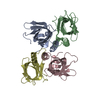
 Assembly
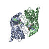
 unidentified baculovirus
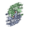
 Mass: 440.512 Da / Num. of mol.: 2 / Source method: isolated from a natural source / Formula: C23H24N2O5S
 Mass: 18.015 Da / Num. of mol.: 30 / Source method: isolated from a natural source / Formula: H2O
 Sample preparation
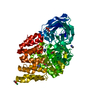
 / Beamline: X06SA / Wavelength: 1 Å
 Processing