登録構造単位
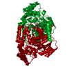
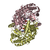
A: Ornithine aminotransferase, mitochondrial
B: Ornithine aminotransferase, mitochondrial
C: Ornithine aminotransferase, mitochondrial
ヘテロ分子 概要 構成要素の詳細
分子量 (理論値) 分子数 合計 (水以外) 138,686 6 ポリマ- 137,945 3 非ポリマー 741 3 水 21,618 1200
1
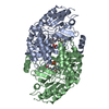
A: Ornithine aminotransferase, mitochondrial
ヘテロ分子
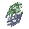
A: Ornithine aminotransferase, mitochondrial
ヘテロ分子 概要 構成要素の詳細 対称操作
分子量 (理論値) 分子数 合計 (水以外) 92,458 4 ポリマ- 91,963 2 非ポリマー 494 2 水 36 2
タイプ 名称 対称操作 数 identity operation 1_555 x,y,z 1 crystal symmetry operation 6_566 x,x-y+1,-z+1 1
2
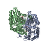
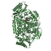
B: Ornithine aminotransferase, mitochondrial
ヘテロ分子
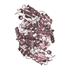
B: Ornithine aminotransferase, mitochondrial
ヘテロ分子 概要 構成要素の詳細 対称操作
分子量 (理論値) 分子数 合計 (水以外) 92,458 4 ポリマ- 91,963 2 非ポリマー 494 2 水 36 2
タイプ 名称 対称操作 数 identity operation 1_555 x,y,z 1 crystal symmetry operation 4_555 -y,-x,-z+2/3 1
3
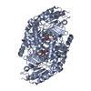
C: Ornithine aminotransferase, mitochondrial
ヘテロ分子
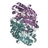
C: Ornithine aminotransferase, mitochondrial
ヘテロ分子 概要 構成要素の詳細 対称操作
分子量 (理論値) 分子数 合計 (水以外) 92,458 4 ポリマ- 91,963 2 非ポリマー 494 2 水 36 2
タイプ 名称 対称操作 数 identity operation 1_555 x,y,z 1 crystal symmetry operation 5_555 -x+y,y,-z+1/3 1
単位格子 Length a, b, c (Å) 193.252, 193.252, 57.211 Angle α, β, γ (deg.) 90.000, 90.000, 120.000 Int Tables number 151 Space group name H-M P31 12
Components on special symmetry positions ID モデル 要素 1 1 A -652-HOH
2 1 A -765-HOH
3 1 A -826-HOH
4 1 A -953-HOH
5 1 B -818-HOH
6 1 B -880-HOH
7 1 B -957-HOH
8 1 B -1000-HOH
9 1 C -654-HOH
10 1 C -734-HOH
11 1 C -793-HOH
非結晶学的対称性 (NCS) NCSドメイン ID Ens-ID 詳細 (eV)1 1 A2 1 B1 2 A2 2 C1 3 B2 3 C
NCSドメイン領域 Component-ID / Beg auth comp-ID / Beg label comp-ID / End auth comp-ID / End label comp-ID / Refine code / Auth seq-ID / Label seq-ID
Dom-ID Ens-ID Auth asym-ID Label asym-ID 1 1 AA2 1 BB1 2 AA2 2 CC1 3 BB2 3 CC
NCSアンサンブル NCS oper ID Code Matrix ベクター 1 given(1), (1), (1)2 given(-0.499299, 0.866429, -0.00066), (-0.86643, -0.499299, 0.000822), (0.000382, 0.000982, 0.999999)-96.585899, 55.760311, 9.536733 given(1), (1), (1)4 given(-0.499487, -0.866315, -0.003167), (0.866309, -0.499496, 0.003408), (-0.004535, -0.001042, 0.999989)0.04461, 111.50779, 19.1585015 given(1), (1), (1)6 given(-0.501118, 0.865379, -0.001003), (-0.865373, -0.501118, -0.003257), (-0.003321, -0.000764, 0.999994)-96.544853, 55.889751, 9.59701
 ムービー
ムービー コントローラー


 データを開く
 基本情報
基本情報 要素
要素 キーワード
キーワード 機能・相同性情報
 Homo sapiens (ヒト)
 X線回折 /
  データ登録者
 イタリア, 2件
  引用
引用 構造の表示
構造の表示 ダウンロードとリンク
 その他のダウンロード
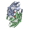


 PDBj
PDBj 集合体
集合体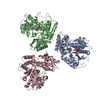


 分子量: 247.142 Da / 分子数: 3 / 由来タイプ: 合成 / 式: C8H10NO6P
 分子量: 18.015 Da / 分子数: 1200 / 由来タイプ: 天然 / 式: H2O
 試料調製
試料調製 解析
解析