 Movie
Movie Controller

[English] 日本語
 Yorodumi
- PDB-1gbn: HUMAN ORNITHINE AMINOTRANSFERASE COMPLEXED WITH THE NEUROTOXIN GA... -
+ Open data

- Basic information
| Entry | Database: PDB / ID: 1gbn | ||||||
|---|---|---|---|---|---|---|---|
| Title | HUMAN ORNITHINE AMINOTRANSFERASE COMPLEXED WITH THE NEUROTOXIN GABACULINE | ||||||
 Components Components | ORNITHINE AMINOTRANSFERASE | ||||||
 Keywords Keywords | TRANSFERASE / ORNITHINE AMINOTRANSFERASE / UREA CYCLE / PYRIDOXAL-5'-PHOSPHATE | ||||||
| Function / homology |  Function and homology information Function and homology information: / ornithine aminotransferase / L-ornithine transaminase activity / : / Glutamate and glutamine metabolism / L-proline biosynthetic process / visual perception / pyridoxal phosphate binding / mitochondrial matrix / mitochondrion ...: / ornithine aminotransferase / L-ornithine transaminase activity / : / Glutamate and glutamine metabolism / L-proline biosynthetic process / visual perception / pyridoxal phosphate binding / mitochondrial matrix / mitochondrion / identical protein binding / cytoplasm Similarity search - Function | ||||||
| Biological species |  Homo sapiens (human) Homo sapiens (human) | ||||||
| Method |  X-RAY DIFFRACTION / SYNCHROTRON / MOLECULAR REPLACEMENT / Resolution: 2.3 Å X-RAY DIFFRACTION / SYNCHROTRON / MOLECULAR REPLACEMENT / Resolution: 2.3 Å | ||||||
 Authors Authors | Shah, S.A. / Shen, B.W. / Brunger, A.T. | ||||||
 Citation Citation | Journal: Structure / Year: 1997 Title: Human ornithine aminotransferase complexed with L-canaline and gabaculine: structural basis for substrate recognition. Authors: Shah, S.A. / Shen, B.W. / Brunger, A.T. | ||||||
| History |
|
- Structure visualization
| Structure viewer | Molecule: MolmilJmol/JSmol |
|---|
- Downloads & links
-Download
| PDBx/mmCIF format | 1gbn.cif.gz | 288.7 KB | Display | PDBx/mmCIF format |
|---|---|---|---|---|
| PDB format | pdb1gbn.ent.gz | 232.9 KB | Display | PDB format |
| PDBx/mmJSON format | 1gbn.json.gz | Tree view | PDBx/mmJSON format | |
| Others |  Other downloads Other downloads |
-Validation report
| Arichive directory | https://data.pdbj.org/pub/pdb/validation_reports/gb/1gbnftp://data.pdbj.org/pub/pdb/validation_reports/gb/1gbn | HTTPS FTP |
|---|
-Related structure data
| Related structure data |  2canC 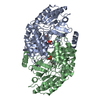 1oatS S: Starting model for refinement C: citing same article ( |
|---|---|
| Similar structure data |


-Links
 PDBj
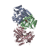
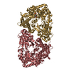
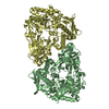
PDBj- Assembly
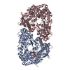
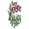
| Deposited unit | 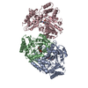
| ||||||||||||
|---|---|---|---|---|---|---|---|---|---|---|---|---|---|
| 1 |
| ||||||||||||
| 2 | 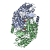
| ||||||||||||
| 3 | 
| ||||||||||||
| 4 | 
| ||||||||||||
| Unit cell |
| ||||||||||||
| Noncrystallographic symmetry (NCS) | NCS oper:
|
-Components
| #1: Protein | Mass: 44706.156 Da / Num. of mol.: 3 Source method: isolated from a genetically manipulated source Details: BOUND INHIBITOR GABACULINE / Source: (gene. exp.) Homo sapiens (human) / Production host:  #2: Chemical | ChemComp-GAB / | 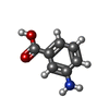  Mass: 137.136 Da / Num. of mol.: 1 / Source method: obtained synthetically / Formula: C7H7NO2 Mass: 137.136 Da / Num. of mol.: 1 / Source method: obtained synthetically / Formula: C7H7NO2#3: Chemical |   Mass: 247.142 Da / Num. of mol.: 3 / Source method: obtained synthetically / Formula: C8H10NO6P Mass: 247.142 Da / Num. of mol.: 3 / Source method: obtained synthetically / Formula: C8H10NO6P#4: Chemical | 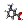  Mass: 141.168 Da / Num. of mol.: 2 / Source method: obtained synthetically / Formula: C7H11NO2 Mass: 141.168 Da / Num. of mol.: 2 / Source method: obtained synthetically / Formula: C7H11NO2#5: Water | ChemComp-HOH / |  Mass: 18.015 Da / Num. of mol.: 329 / Source method: isolated from a natural source / Formula: H2O Mass: 18.015 Da / Num. of mol.: 329 / Source method: isolated from a natural source / Formula: H2O |
|---|
-Experimental details
-Experiment
| Experiment | Method: X-RAY DIFFRACTION / Number of used crystals: 1 |
|---|
- Sample preparation
| Crystal | Density Matthews: 2.64 Å3/Da / Density % sol: 50.1 % | ||||||||||||||||||||||||||||||||||||
|---|---|---|---|---|---|---|---|---|---|---|---|---|---|---|---|---|---|---|---|---|---|---|---|---|---|---|---|---|---|---|---|---|---|---|---|---|---|
| Crystal grow | pH: 6.5 / Details: pH 6.5 | ||||||||||||||||||||||||||||||||||||
| Crystal grow | *PLUS pH: 7.8 / Method: vapor diffusion, hanging drop | ||||||||||||||||||||||||||||||||||||
| Components of the solutions | *PLUS
|
-Data collection
| Diffraction | Mean temperature: 110 K |
|---|---|
| Diffraction source | Source: SYNCHROTRON / Site: CHESS  / Beamline: A1 / Wavelength: 0.9417 / Beamline: A1 / Wavelength: 0.9417 |
| Detector | Type: PRINCETON 2K / Detector: CCD / Date: Oct 10, 1995 / Details: DUEL SLITS |
| Radiation | Monochromator: CRYSTAL / Monochromatic (M) / Laue (L): M / Scattering type: x-ray |
| Radiation wavelength | Wavelength: 0.9417 Å / Relative weight: 1 |
| Reflection | Resolution: 2.3→50 Å / Num. obs: 57010 / % possible obs: 90 % / Observed criterion σ(I): 2 / Redundancy: 4.6 % / Biso Wilson estimate: 14.3 Å2 / Rmerge(I) obs: 0.064 / Rsym value: 0.064 / Net I/σ(I): 16.2 |
| Reflection shell | Resolution: 2.3→2.38 Å / Redundancy: 4.6 % / Rmerge(I) obs: 0.17 / Mean I/σ(I) obs: 5.9 / Rsym value: 0.17 / % possible all: 80.1 |
| Reflection | *PLUS % possible obs: 89.9 % |
| Reflection shell | *PLUS Lowest resolution: 2.4 Å / % possible obs: 80.1 % / Redundancy: 2.6 % / Rmerge(I) obs: 0.171 |
- Processing
| Software |
| ||||||||||||||||||||||||||||||||||||||||||||||||||||||||||||||||||||||||||||||||
|---|---|---|---|---|---|---|---|---|---|---|---|---|---|---|---|---|---|---|---|---|---|---|---|---|---|---|---|---|---|---|---|---|---|---|---|---|---|---|---|---|---|---|---|---|---|---|---|---|---|---|---|---|---|---|---|---|---|---|---|---|---|---|---|---|---|---|---|---|---|---|---|---|---|---|---|---|---|---|---|---|---|
| Refinement | Method to determine structure: MOLECULAR REPLACEMENT Starting model: PDB ENTRY 1OAT Resolution: 2.3→50 Å / Rfactor Rfree error: 0.003 / Data cutoff high absF: 470426.53 / Data cutoff low absF: 0.0001 / Isotropic thermal model: RESTRAINED / Cross valid method: THROUGHOUT / σ(F): 2 / Details: BULK SOLVENT MODEL USED
| ||||||||||||||||||||||||||||||||||||||||||||||||||||||||||||||||||||||||||||||||
| Displacement parameters | Biso mean: 22 Å2
| ||||||||||||||||||||||||||||||||||||||||||||||||||||||||||||||||||||||||||||||||
| Refine analyze |
| ||||||||||||||||||||||||||||||||||||||||||||||||||||||||||||||||||||||||||||||||
| Refinement step | Cycle: LAST / Resolution: 2.3→50 Å
| ||||||||||||||||||||||||||||||||||||||||||||||||||||||||||||||||||||||||||||||||
| Refine LS restraints |
| ||||||||||||||||||||||||||||||||||||||||||||||||||||||||||||||||||||||||||||||||
| Refine LS restraints NCS | Rms dev Biso : 1.618 Å2 / Rms dev position: 0.1647 Å | ||||||||||||||||||||||||||||||||||||||||||||||||||||||||||||||||||||||||||||||||
| LS refinement shell | Resolution: 2.3→2.44 Å / Rfactor Rfree error: 0.011 / Total num. of bins used: 6
| ||||||||||||||||||||||||||||||||||||||||||||||||||||||||||||||||||||||||||||||||
| Xplor file |
| ||||||||||||||||||||||||||||||||||||||||||||||||||||||||||||||||||||||||||||||||
| Software | *PLUS Name: X-PLOR / Version: 3.851 / Classification: refinement | ||||||||||||||||||||||||||||||||||||||||||||||||||||||||||||||||||||||||||||||||
| Refinement | *PLUS Num. reflection obs: 52218 / Rfactor obs: 0.206 | ||||||||||||||||||||||||||||||||||||||||||||||||||||||||||||||||||||||||||||||||
| Solvent computation | *PLUS | ||||||||||||||||||||||||||||||||||||||||||||||||||||||||||||||||||||||||||||||||
| Displacement parameters | *PLUS | ||||||||||||||||||||||||||||||||||||||||||||||||||||||||||||||||||||||||||||||||
| Refine LS restraints | *PLUS
| ||||||||||||||||||||||||||||||||||||||||||||||||||||||||||||||||||||||||||||||||
| LS refinement shell | *PLUS Highest resolution: 2.3 Å / Lowest resolution: 2.4 Å / Num. reflection Rfree: 70 / % reflection Rfree: 10 % / Num. reflection Rwork: 5123 |
