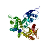
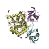
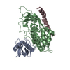
Resolution: 1.9→39.403 Å / SU ML: 0.28 / Isotropic thermal model: isotropic / Cross valid method: THROUGHOUT / σ(F): 0 / Phase error: 21.41 / Stereochemistry target values: ML Details: AUTHORS DO NOT OBSERVE RESIDUES 295-514 (THE ALPHA-HELICAL DOMAIN) IN THE CRYSTAL STRUCTURE. THEY SUSPECT LIMITED PROTEOLYSIS DURING CRYSTALLISATION
Rfactor
Num. reflection
% reflection
Selection details
Rfree
0.218
3430
5.07 %
RANDOM
Rwork
0.1807
-
-
-
all
0.1826
67702
-
-
obs
0.1826
67702
95.75 %
-
Solvent computation
Shrinkage radii: 0.83 Å / VDW probe radii: 1.1 Å / Solvent model: FLAT BULK SOLVENT MODEL / Bsol: 38.725 Å2 / ksol: 0.319 e/Å3
Displacement parameters
Baniso -1
Baniso -2
Baniso -3
1-
3.2102 Å2
-0 Å2
0.7326 Å2
2-
-
-5.395 Å2
-0 Å2
3-
-
-
2.1848 Å2
Refinement step
Cycle: LAST / Resolution: 1.9→39.403 Å
Protein
Nucleic acid
Ligand
Solvent
Total
Num. atoms
6144
0
0
564
6708
Refine LS restraints
Refine-ID
Type
Dev ideal
Number
X-RAY DIFFRACTION
f_bond_d
0.005
6330
X-RAY DIFFRACTION
f_angle_d
0.894
8521
X-RAY DIFFRACTION
f_dihedral_angle_d
13.675
2481
X-RAY DIFFRACTION
f_chiral_restr
0.066
897
X-RAY DIFFRACTION
f_plane_restr
0.003
1110
LS refinement shell
Resolution (Å)
Rfactor Rfree
Num. reflection Rfree
Rfactor Rwork
Num. reflection Rwork
Refine-ID
% reflection obs (%)
1.9-1.9679
0.3327
324
0.2768
6122
X-RAY DIFFRACTION
92
1.9679-2.0467
0.2703
329
0.2099
6122
X-RAY DIFFRACTION
91
2.0467-2.1399
0.2424
342
0.1951
6046
X-RAY DIFFRACTION
91
2.1399-2.2527
0.2275
319
0.181
6041
X-RAY DIFFRACTION
90
2.2527-2.3938
0.2452
326
0.1921
6446
X-RAY DIFFRACTION
96
2.3938-2.5786
0.2436
351
0.1913
6704
X-RAY DIFFRACTION
100
2.5786-2.838
0.2358
368
0.1836
6712
X-RAY DIFFRACTION
100
2.838-3.2485
0.2357
359
0.1881
6692
X-RAY DIFFRACTION
100
3.2485-4.0921
0.1906
354
0.1702
6707
X-RAY DIFFRACTION
99
4.0921-39.411
0.1795
358
0.1579
6680
X-RAY DIFFRACTION
98
Refinement TLS params.
Method: refined / Refine-ID: X-RAY DIFFRACTION
ID
L11 (°2)
L12 (°2)
L13 (°2)
L22 (°2)
L23 (°2)
L33 (°2)
S11 (Å °)
S12 (Å °)
S13 (Å °)
S21 (Å °)
S22 (Å °)
S23 (Å °)
S31 (Å °)
S32 (Å °)
S33 (Å °)
T11 (Å2)
T12 (Å2)
T13 (Å2)
T22 (Å2)
T23 (Å2)
T33 (Å2)
Origin x (Å)
Origin y (Å)
Origin z (Å)
1
1.4802
-0.0933
0.0377
1.9063
1.9581
2.233
0.2005
0.0632
0.3232
-0.2205
-0.1805
0.0463
-0.4488
-0.1276
-0.0402
0.3482
0.0373
0.0222
0.1016
-0.0005
0.123
21.5143
42.5769
11.9242
2
1.185
-0.838
-0.2756
2.3585
-0.7114
1.2037
-0.0631
-0.0983
0.1584
0.2554
-0.0283
-0.4127
-0.3195
0.0822
0.1063
0.2604
-0.02
-0.0982
0.1269
-0.0334
0.2031
33.766
18.2295
22.9778
3
1.2024
-0.2956
0.4079
1.8478
0.2042
1.992
-0.0581
0.1316
-0.0158
-0.289
-0.0393
0.4081
-0.3073
-0.3925
0.1045
0.2478
0.0692
-0.1494
0.2167
-0.0421
0.258
6.4709
21.108
8.0997
4
0.4831
-0.5033
-0.2664
0.6891
0.6892
1.1683
-0.0806
0.044
-0.1904
0.0309
-0.0928
-0.2003
0.3165
0.0525
0.0552
0.2502
0.0156
-0.058
0.1513
-0.0363
0.2642
31.129
4.8774
20.0642
5
1.1196
-1.6696
-0.2312
4.1312
-1.1616
1.4374
-0.3085
-0.3649
-0.3938
0.8783
0.2147
0.3951
0.2881
-0.2689
0.0921
0.2996
0.0701
0.0557
0.2217
0.0283
0.2583
16.499
5.8876
28.9678
6
1.6343
-1.2547
0.7304
1.3473
0.1209
2.5967
-0.0014
0.5181
-0.6061
-0.5106
0.0808
0.6083
0.138
-0.0043
-0.1158
0.3328
-0.0103
-0.029
0.2033
-0.0735
0.3055
14.4243
6.5466
10.1315
7
3.6236
0.9336
2.5438
0.3117
0.4601
2.7785
-0.1801
0.3346
-0.7023
-0.5747
0.0346
0.322
0.3266
-0.4935
0.097
0.315
-0.0856
-0.2001
0.3361
-0.0553
0.5542
-1.0828
9.4184
7.5843
8
2.3561
-0.738
-1.0657
1.2098
1.2952
3.8662
-0.1631
0.1389
-0.5197
0.3181
-0.0731
0.2503
0.7484
-0.3018
0.2435
0.2464
-0.0301
0.0655
0.1063
-0.021
0.2772
5.362
14.8661
45.662
9
1.862
-0.3195
0.839
1.393
0.1982
2.6569
-0.0045
0.1517
-0.0778
0.1985
0.0014
-0.1976
0.0517
0.4911
0.005
0.1169
0.021
-0.0334
0.2253
0.0009
0.1446
25.0729
35.0833
48.6969
10
1.1212
-0.3527
0.3177
2.2161
-0.17
1.9534
-0.0829
0.1259
-0.1513
-0.1037
0.0017
0.4128
0.062
-0.167
0.0701
0.0708
-0.0172
-0.0275
0.1728
-0.0339
0.2648
-4.3989
37.0646
38.2239
11
3.4259
-0.1795
0.2009
1.0306
0.0037
0.788
-0.1239
-0.2619
0.8917
0.4215
0.1186
-0.0149
-0.3735
0.2595
0.0764
0.254
-0.0743
-0.0465
0.2081
0.0284
0.2583
21.55
49.0598
49.5302
12
1.0023
0.1525
-0.4951
0.9551
0.3492
1.762
0.0606
0.3767
0.0216
-0.4819
-0.0632
-0.2707
-0.4761
0.375
0.0256
0.1823
-0.071
0.0228
0.2412
0.0488
0.1207
19.165
49.2258
32.9716
13
3.1945
-0.8875
1.904
0.7752
-0.4091
2.1044
-0.1362
-0.1906
0.2828
0.0169
-0.0426
0.0176
-0.0039
-0.1849
0.1808
0.1684
0.0043
0.0137
0.1446
0.0004
0.2056
4.1012
50.1856
43.7728
14
3.7295
1.8971
-2.5386
0.9655
-1.32
5.1051
0.0753
-0.1498
0.7308
-0.2876
0.1252
0.819
-0.7447
-0.318
0.1616
0.1851
0.0605
-0.1588
0.1959
0.0569
0.4093
-8.2221
49.6676
33.7954
Refinement TLS group
ID
Refine-ID
Refine TLS-ID
Selection details
Auth asym-ID
Auth seq-ID
1
X-RAY DIFFRACTION
1
( CHAINAANDRESID1:85 )
A
1 - 85
2
X-RAY DIFFRACTION
2
( CHAINAANDRESID86:200 )
A
86 - 200
3
X-RAY DIFFRACTION
3
( CHAINAANDRESID201:297 )
A
201 - 297
4
X-RAY DIFFRACTION
4
( CHAINAANDRESID516:542 )
A
516 - 542
5
X-RAY DIFFRACTION
5
( CHAINAANDRESID543:562 )
A
543 - 562
6
X-RAY DIFFRACTION
6
( CHAINAANDRESID563:573 )
A
563 - 573
7
X-RAY DIFFRACTION
7
( CHAINAANDRESID574:586 )
A
574 - 586
8
X-RAY DIFFRACTION
8
( CHAINBANDRESID3:85 )
B
3 - 85
9
X-RAY DIFFRACTION
9
( CHAINBANDRESID86:200 )
B
86 - 200
10
X-RAY DIFFRACTION
10
( CHAINBANDRESID201:297 )
B
201 - 297
11
X-RAY DIFFRACTION
11
( CHAINBANDRESID515:542 )
B
515 - 542
12
X-RAY DIFFRACTION
12
( CHAINBANDRESID543:562 )
B
543 - 562
13
X-RAY DIFFRACTION
13
( CHAINBANDRESID563:573 )
B
563 - 573
14
X-RAY DIFFRACTION
14
( CHAINBANDRESID574:586 )
B
574 - 586
+
About Yorodumi
-
News
-
Feb 9, 2022. New format data for meta-information of EMDB entries
New format data for meta-information of EMDB entries
Version 3 of the EMDB header file is now the official format.
The previous official version 1.9 will be removed from the archive.
In the structure databanks used in Yorodumi, some data are registered as the other names, "COVID-19 virus" and "2019-nCoV". Here are the details of the virus and the list of structure data.
Jan 31, 2019. EMDB accession codes are about to change! (news from PDBe EMDB page)
EMDB accession codes are about to change! (news from PDBe EMDB page)
The allocation of 4 digits for EMDB accession codes will soon come to an end. Whilst these codes will remain in use, new EMDB accession codes will include an additional digit and will expand incrementally as the available range of codes is exhausted. The current 4-digit format prefixed with “EMD-” (i.e. EMD-XXXX) will advance to a 5-digit format (i.e. EMD-XXXXX), and so on. It is currently estimated that the 4-digit codes will be depleted around Spring 2019, at which point the 5-digit format will come into force.
The EM Navigator/Yorodumi systems omit the EMD- prefix.
Related info.:Q: What is EMD? / ID/Accession-code notation in Yorodumi/EM Navigator
Yorodumi is a browser for structure data from EMDB, PDB, SASBDB, etc.
This page is also the successor to EM Navigator detail page, and also detail information page/front-end page for Omokage search.
The word "yorodu" (or yorozu) is an old Japanese word meaning "ten thousand". "mi" (miru) is to see.
Related info.:EMDB / PDB / SASBDB / Comparison of 3 databanks / Yorodumi Search / Aug 31, 2016. New EM Navigator & Yorodumi / Yorodumi Papers / Jmol/JSmol / Function and homology information / Changes in new EM Navigator and Yorodumi
 Movie
Movie Controller


 Open data
 Basic information
 Components
 Keywords
 Function and homology information
 Homo sapiens (human)
 X-RAY DIFFRACTION /
  Authors
 Citation
 Structure visualization
 Downloads & links
 Other downloads


 PDBj
PDBj


 Assembly
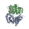


 Mass: 18.015 Da / Num. of mol.: 564 / Source method: isolated from a natural source / Formula: H2O
 Sample preparation
 / Beamline: MX2 / Wavelength: 0.954 Å
 Processing