 Movie
Movie Controller

[English] 日本語
 Yorodumi
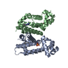
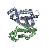
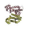
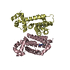
- PDB-6ko9: Crystal structure of the Gefitinib Intermediate 1 bound RamR dete... -
+ Open data

- Basic information
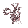
| Entry | Database: PDB / ID: 6ko9 | ||||||
|---|---|---|---|---|---|---|---|
| Title | Crystal structure of the Gefitinib Intermediate 1 bound RamR determined with XtaLAB Synergy | ||||||
 Components Components | Putative regulatory protein | ||||||
 Keywords Keywords | DNA BINDING PROTEIN / multisite binding pocket / HTH-MOTIF | ||||||
| Function / homology |  Function and homology information Function and homology information | ||||||
| Biological species |  Salmonella enterica subsp. enterica serovar Typhimurium str. 14028S (bacteria) Salmonella enterica subsp. enterica serovar Typhimurium str. 14028S (bacteria) | ||||||
| Method |  X-RAY DIFFRACTION / MOLECULAR REPLACEMENT / molecular replacement / Resolution: 2.2 Å X-RAY DIFFRACTION / MOLECULAR REPLACEMENT / molecular replacement / Resolution: 2.2 Å | ||||||
| Model details | framework as crystalline sponge method | ||||||
 Authors Authors | Matsumoto, T. / Nakashima, R. / Yamano, A. / Nishino, K. | ||||||
 Citation Citation | Journal: Biochem.Biophys.Res.Commun. / Year: 2019 Title: Development of a structure determination method using a multidrug-resistance regulator protein as a framework. Authors: Matsumoto, T. / Nakashima, R. / Yamano, A. / Nishino, K. | ||||||
| History |
|
- Structure visualization
| Structure viewer | Molecule: MolmilJmol/JSmol |
|---|
- Downloads & links
-Download
| PDBx/mmCIF format | 6ko9.cif.gz | 176.1 KB | Display | PDBx/mmCIF format |
|---|---|---|---|---|
| PDB format | pdb6ko9.ent.gz | 138.9 KB | Display | PDB format |
| PDBx/mmJSON format | 6ko9.json.gz | Tree view | PDBx/mmJSON format | |
| Others |  Other downloads Other downloads |
-Validation report
| Arichive directory | https://data.pdbj.org/pub/pdb/validation_reports/ko/6ko9ftp://data.pdbj.org/pub/pdb/validation_reports/ko/6ko9 | HTTPS FTP |
|---|
-Related structure data
| Related structure data |  6ko7C  6ko8C  3vvyS S: Starting model for refinement C: citing same article ( |
|---|---|
| Similar structure data |


-Links
 PDBj
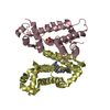
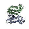
PDBj- Assembly
| Deposited unit | 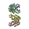
| ||||||||
|---|---|---|---|---|---|---|---|---|---|
| 1 | 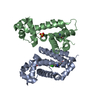
| ||||||||
| 2 | 
| ||||||||
| Unit cell |
|
-Components
| #1: Protein | Mass: 22044.262 Da / Num. of mol.: 4 Source method: isolated from a genetically manipulated source Source: (gene. exp.) Salmonella enterica subsp. enterica serovar Typhimurium str. 14028S (bacteria)Strain: 14028s / SGSC 2262 / Gene: STM14_0676 / Plasmid: PET DUET / Production host: #2: Chemical | ChemComp-XZ1 / 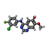  Mass: 319.718 Da / Num. of mol.: 4 / Source method: obtained synthetically / Formula: C15H11ClFN3O2 / Feature type: SUBJECT OF INVESTIGATION Mass: 319.718 Da / Num. of mol.: 4 / Source method: obtained synthetically / Formula: C15H11ClFN3O2 / Feature type: SUBJECT OF INVESTIGATION#3: Chemical | ChemComp-SO4 /   Mass: 96.063 Da / Num. of mol.: 5 / Source method: obtained synthetically / Formula: SO4 Mass: 96.063 Da / Num. of mol.: 5 / Source method: obtained synthetically / Formula: SO4#4: Water | ChemComp-HOH / |  Mass: 18.015 Da / Num. of mol.: 662 / Source method: isolated from a natural source / Formula: H2O Mass: 18.015 Da / Num. of mol.: 662 / Source method: isolated from a natural source / Formula: H2OHas ligand of interest | Y | |
|---|
-Experimental details
-Experiment
| Experiment | Method: X-RAY DIFFRACTION / Number of used crystals: 1 |
|---|
- Sample preparation
| Crystal | Density Matthews: 2.51 Å3/Da / Density % sol: 51.01 % / Mosaicity: 0.99 ° |
|---|---|
| Crystal grow | Temperature: 298 K / Method: vapor diffusion, sitting drop / pH: 5.6 / Details: PEG3350, ammonium sulfate, sodium Citrate |
-Data collection
| Diffraction | Mean temperature: 100 K / Serial crystal experiment: N | ||||||||||||||||||||||||||||||
|---|---|---|---|---|---|---|---|---|---|---|---|---|---|---|---|---|---|---|---|---|---|---|---|---|---|---|---|---|---|---|---|
| Diffraction source | Source: ROTATING ANODE / Type: RIGAKU FR-X / Wavelength: 1.5406 Å | ||||||||||||||||||||||||||||||
| Detector | Type: RIGAKU HyPic-6000HE / Detector: PIXEL / Date: Nov 25, 2016 | ||||||||||||||||||||||||||||||
| Radiation | Monochromator: multi-layer mirror optics / Protocol: SINGLE WAVELENGTH / Monochromatic (M) / Laue (L): M / Scattering type: x-ray | ||||||||||||||||||||||||||||||
| Radiation wavelength | Wavelength: 1.5406 Å / Relative weight: 1 | ||||||||||||||||||||||||||||||
| Reflection | Resolution: 2.2→18.44 Å / Num. obs: 40974 / % possible obs: 99.3 % / Redundancy: 3.4 % / CC1/2: 0.992 / Rmerge(I) obs: 0.106 / Rpim(I) all: 0.062 / Rrim(I) all: 0.123 / Net I/σ(I): 6.2 / Num. measured all: 138507 / Scaling rejects: 554 | ||||||||||||||||||||||||||||||
| Reflection shell | Diffraction-ID: 1
|
-Phasing
| Phasing | Method: molecular replacement |
|---|
- Processing
| Software |
| ||||||||||||||||||||||||||||||||||||||||||||||||||||||||||||
|---|---|---|---|---|---|---|---|---|---|---|---|---|---|---|---|---|---|---|---|---|---|---|---|---|---|---|---|---|---|---|---|---|---|---|---|---|---|---|---|---|---|---|---|---|---|---|---|---|---|---|---|---|---|---|---|---|---|---|---|---|---|
| Refinement | Method to determine structure: MOLECULAR REPLACEMENT Starting model: 3VVY Resolution: 2.2→18.44 Å / Cor.coef. Fo:Fc: 0.953 / Cor.coef. Fo:Fc free: 0.914 / SU B: 11.223 / SU ML: 0.265 / Cross valid method: THROUGHOUT / σ(F): 0 / ESU R: 0.364 / ESU R Free: 0.278 / Stereochemistry target values: MAXIMUM LIKELIHOOD Details: HYDROGENS HAVE BEEN ADDED IN THE RIDING POSITIONS U VALUES : REFINED INDIVIDUALLY
| ||||||||||||||||||||||||||||||||||||||||||||||||||||||||||||
| Solvent computation | Ion probe radii: 0.8 Å / Shrinkage radii: 0.8 Å / VDW probe radii: 1.2 Å / Solvent model: MASK | ||||||||||||||||||||||||||||||||||||||||||||||||||||||||||||
| Displacement parameters | Biso max: 327.2 Å2 / Biso mean: 57.627 Å2 / Biso min: 27.47 Å2
| ||||||||||||||||||||||||||||||||||||||||||||||||||||||||||||
| Refinement step | Cycle: final / Resolution: 2.2→18.44 Å
| ||||||||||||||||||||||||||||||||||||||||||||||||||||||||||||
| Refine LS restraints |
| ||||||||||||||||||||||||||||||||||||||||||||||||||||||||||||
| LS refinement shell | Resolution: 2.2→2.257 Å / Rfactor Rfree error: 0 / Total num. of bins used: 20
|
