 Movie
Movie Controller

[English] 日本語
 Yorodumi
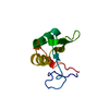
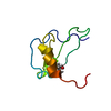
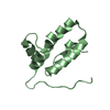
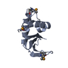
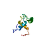
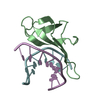
- PDB-4r55: The crystal structure of a Cren7 mutant protein GR and dsDNA complex -
+ Open data

- Basic information
| Entry | Database: PDB / ID: 4r55 | ||||||
|---|---|---|---|---|---|---|---|
| Title | The crystal structure of a Cren7 mutant protein GR and dsDNA complex | ||||||
 Components Components |
| ||||||
 Keywords Keywords | DNA BINDING PROTEIN/DNA / BETA-SHEET / DNA BINDING / DNA BINDING PROTEIN-DNA complex | ||||||
| Function / homology |  Function and homology information Function and homology information | ||||||
| Biological species |   Sulfolobus solfataricus P2 (archaea) Sulfolobus solfataricus P2 (archaea)synthetic (others) | ||||||
| Method |  X-RAY DIFFRACTION / MOLECULAR REPLACEMENT / Resolution: 1.8 Å X-RAY DIFFRACTION / MOLECULAR REPLACEMENT / Resolution: 1.8 Å | ||||||
 Authors Authors | Zhang, Z.F. / Gong, Y. / Chen, Y.Y. / Li, H.B. / Huang, L. | ||||||
 Citation Citation | Journal: Extremophiles / Year: 2015 Title: Insights into the interaction between Cren7 and DNA: the role of loop beta 3-beta 4 Authors: Zhang, Z.F. / Gong, Y. / Chen, Y.Y. / Li, H.B. / Huang, L. | ||||||
| History |
|
- Structure visualization
| Structure viewer | Molecule: MolmilJmol/JSmol |
|---|
- Downloads & links
-Download
| PDBx/mmCIF format | 4r55.cif.gz | 34.8 KB | Display | PDBx/mmCIF format |
|---|---|---|---|---|
| PDB format | pdb4r55.ent.gz | 20.8 KB | Display | PDB format |
| PDBx/mmJSON format | 4r55.json.gz | Tree view | PDBx/mmJSON format | |
| Others |  Other downloads Other downloads |
-Validation report
| Summary document | 4r55_validation.pdf.gz | 405.9 KB | Display | wwPDB validaton report |
|---|---|---|---|---|
| Full document | 4r55_full_validation.pdf.gz | 405.9 KB | Display | |
| Data in XML | 4r55_validation.xml.gz | 5.5 KB | Display | |
| Data in CIF | 4r55_validation.cif.gz | 7 KB | Display | |
| Arichive directory | https://data.pdbj.org/pub/pdb/validation_reports/r5/4r55ftp://data.pdbj.org/pub/pdb/validation_reports/r5/4r55 | HTTPS FTP |
-Related structure data
| Related structure data |  4r56C  3lwhS S: Starting model for refinement C: citing same article ( |
|---|---|
| Similar structure data |


-Links
 PDBj
PDBj- Assembly
| Deposited unit | 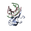
| ||||||||
|---|---|---|---|---|---|---|---|---|---|
| 1 |
| ||||||||
| Unit cell |
|
-Components
| #1: Protein | Mass: 6194.311 Da / Num. of mol.: 1 / Mutation: DELETION Source method: isolated from a genetically manipulated source Source: (gene. exp.) Sulfolobus solfataricus P2 (archaea) / Gene: creN7, SSO6901 / Plasmid: pET30a / Production host:  | ||
|---|---|---|---|
| #2: DNA chain | Mass: 2426.617 Da / Num. of mol.: 2 / Source method: obtained synthetically / Source: (synth.) synthetic (others) #3: Water | ChemComp-HOH / |  Mass: 18.015 Da / Num. of mol.: 72 / Source method: isolated from a natural source / Formula: H2O Mass: 18.015 Da / Num. of mol.: 72 / Source method: isolated from a natural source / Formula: H2O |
-Experimental details
-Experiment
| Experiment | Method: X-RAY DIFFRACTION / Number of used crystals: 1 |
|---|
- Sample preparation
| Crystal | Density Matthews: 2.18 Å3/Da / Density % sol: 43.61 % |
|---|---|
| Crystal grow | Temperature: 293 K / Method: vapor diffusion, sitting drop / pH: 6.5 Details: 0.2M NH4Ac, 0.01M MgAc2, 0.05M sodium cacodylate trihydrate, 30% PEG 8000, pH 6.5, VAPOR DIFFUSION, SITTING DROP, temperature 293K |
-Data collection
| Diffraction | Mean temperature: 100 K |
|---|---|
| Diffraction source | Source: ROTATING ANODE / Type: RIGAKU MICROMAX-007 HF / Wavelength: 1.5418 Å |
| Detector | Type: RIGAKU RAXIS IV++ / Detector: IMAGE PLATE / Date: May 17, 2013 |
| Radiation | Monochromator: GRAPHITE / Protocol: SINGLE WAVELENGTH / Monochromatic (M) / Laue (L): M / Scattering type: x-ray |
| Radiation wavelength | Wavelength: 1.5418 Å / Relative weight: 1 |
| Reflection | Resolution: 1.8→50 Å / Num. all: 9449 / Num. obs: 9402 / % possible obs: 99.5 % / Observed criterion σ(F): 1 / Observed criterion σ(I): 1 / Redundancy: 6.7 % / Biso Wilson estimate: 12.9 Å2 / Rmerge(I) obs: 0.051 / Net I/σ(I): 40.6 |
| Reflection shell | Resolution: 1.8→1.86 Å / Redundancy: 6.6 % / Rmerge(I) obs: 0.084 / Mean I/σ(I) obs: 25.3 / Num. unique all: 918 / % possible all: 99 |
- Processing
| Software |
| ||||||||||||||||||||||||||||||||||||||||||||||||||||||||||||||||||||||||||||||||||||||||||||||||||||||||||||||||||||||||||||||||||||||||||||||||||||||||||||||||||||||||||||||||||||||
|---|---|---|---|---|---|---|---|---|---|---|---|---|---|---|---|---|---|---|---|---|---|---|---|---|---|---|---|---|---|---|---|---|---|---|---|---|---|---|---|---|---|---|---|---|---|---|---|---|---|---|---|---|---|---|---|---|---|---|---|---|---|---|---|---|---|---|---|---|---|---|---|---|---|---|---|---|---|---|---|---|---|---|---|---|---|---|---|---|---|---|---|---|---|---|---|---|---|---|---|---|---|---|---|---|---|---|---|---|---|---|---|---|---|---|---|---|---|---|---|---|---|---|---|---|---|---|---|---|---|---|---|---|---|---|---|---|---|---|---|---|---|---|---|---|---|---|---|---|---|---|---|---|---|---|---|---|---|---|---|---|---|---|---|---|---|---|---|---|---|---|---|---|---|---|---|---|---|---|---|---|---|---|---|
| Refinement | Method to determine structure: MOLECULAR REPLACEMENT Starting model: 3LWH Resolution: 1.8→17 Å / Cor.coef. Fo:Fc: 0.927 / Cor.coef. Fo:Fc free: 0.893 / SU B: 2.857 / SU ML: 0.092 / Cross valid method: THROUGHOUT / σ(F): 1 / ESU R: 0.152 / ESU R Free: 0.14 / Stereochemistry target values: MAXIMUM LIKELIHOOD
| ||||||||||||||||||||||||||||||||||||||||||||||||||||||||||||||||||||||||||||||||||||||||||||||||||||||||||||||||||||||||||||||||||||||||||||||||||||||||||||||||||||||||||||||||||||||
| Solvent computation | Ion probe radii: 0.8 Å / Shrinkage radii: 0.8 Å / VDW probe radii: 1.2 Å / Solvent model: MASK | ||||||||||||||||||||||||||||||||||||||||||||||||||||||||||||||||||||||||||||||||||||||||||||||||||||||||||||||||||||||||||||||||||||||||||||||||||||||||||||||||||||||||||||||||||||||
| Displacement parameters | Biso mean: 12.603 Å2
| ||||||||||||||||||||||||||||||||||||||||||||||||||||||||||||||||||||||||||||||||||||||||||||||||||||||||||||||||||||||||||||||||||||||||||||||||||||||||||||||||||||||||||||||||||||||
| Refinement step | Cycle: LAST / Resolution: 1.8→17 Å
| ||||||||||||||||||||||||||||||||||||||||||||||||||||||||||||||||||||||||||||||||||||||||||||||||||||||||||||||||||||||||||||||||||||||||||||||||||||||||||||||||||||||||||||||||||||||
| Refine LS restraints |
|