- PDB-2eo2: Solution structure of the insertion region (510-573) of FTHFS dom... -
+
Open data
ID or keywords:
Loading...
-
Basic information
Entry
Database: PDB / ID: 2eo2
Title
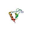
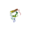
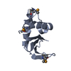
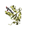
Solution structure of the insertion region (510-573) of FTHFS domain from mouse methylenetetrahydrofolate dehydrogenase (NADP+ dependent) 1-like protein
Components
Adult male hypothalamus cDNA, RIKEN full-length enriched library, clone:A230045M11 product:weakly similar to C1-tetrahydrofolate synthase
Keywords
OXIDOREDUCTASE / Fthfsdc1 / Structural Genomics / NPPSFA / National Project on Protein Structural and Functional Analyses / RIKEN Structural Genomics/Proteomics Initiative / RSGI
Function / homology
Function and homology information
Metabolism of folate and pterines / formate-tetrahydrofolate ligase / formate-tetrahydrofolate ligase activity / embryonic neurocranium morphogenesis / formate metabolic process / formate biosynthetic process / embryonic viscerocranium morphogenesis / methylenetetrahydrofolate dehydrogenase (NADP+) activity / folic acid-containing compound metabolic process / 10-formyltetrahydrofolate biosynthetic process ...Metabolism of folate and pterines / formate-tetrahydrofolate ligase / formate-tetrahydrofolate ligase activity / embryonic neurocranium morphogenesis / formate metabolic process / formate biosynthetic process / embryonic viscerocranium morphogenesis / methylenetetrahydrofolate dehydrogenase (NADP+) activity / folic acid-containing compound metabolic process / 10-formyltetrahydrofolate biosynthetic process / tetrahydrofolate interconversion / neural tube closure / mitochondrial matrix / protein homodimerization activity / mitochondrion / ATP binding / cytosol Similarity search - Function
Conformer selection criteria: structures with the least restraint violations, structures with the lowest energy, target function Conformers calculated total number: 100 / Conformers submitted total number: 20
+
About Yorodumi
-
News
-
Feb 9, 2022. New format data for meta-information of EMDB entries
New format data for meta-information of EMDB entries
Version 3 of the EMDB header file is now the official format.
The previous official version 1.9 will be removed from the archive.
In the structure databanks used in Yorodumi, some data are registered as the other names, "COVID-19 virus" and "2019-nCoV". Here are the details of the virus and the list of structure data.
Jan 31, 2019. EMDB accession codes are about to change! (news from PDBe EMDB page)
EMDB accession codes are about to change! (news from PDBe EMDB page)
The allocation of 4 digits for EMDB accession codes will soon come to an end. Whilst these codes will remain in use, new EMDB accession codes will include an additional digit and will expand incrementally as the available range of codes is exhausted. The current 4-digit format prefixed with “EMD-” (i.e. EMD-XXXX) will advance to a 5-digit format (i.e. EMD-XXXXX), and so on. It is currently estimated that the 4-digit codes will be depleted around Spring 2019, at which point the 5-digit format will come into force.
The EM Navigator/Yorodumi systems omit the EMD- prefix.
Related info.:Q: What is EMD? / ID/Accession-code notation in Yorodumi/EM Navigator
Yorodumi is a browser for structure data from EMDB, PDB, SASBDB, etc.
This page is also the successor to EM Navigator detail page, and also detail information page/front-end page for Omokage search.
The word "yorodu" (or yorozu) is an old Japanese word meaning "ten thousand". "mi" (miru) is to see.
Related info.:EMDB / PDB / SASBDB / Comparison of 3 databanks / Yorodumi Search / Aug 31, 2016. New EM Navigator & Yorodumi / Yorodumi Papers / Jmol/JSmol / Function and homology information / Changes in new EM Navigator and Yorodumi
 Movie
Movie Controller

 Yorodumi
 Open data
 Basic information
 Components
 Keywords
 Function and homology information
 Authors
 Citation
 Structure visualization
 Downloads & links
 Other downloads
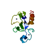


 PDBj
PDBj Assembly
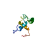
 Sample preparation
 Processing
 NMRPipe