 Movie
Movie Controller

[English] 日本語
 Yorodumi
- PDB-4q4g: Structure of the Resuscitation Promoting Factor Interacting prote... -
+ Open data

- Basic information
| Entry | Database: PDB / ID: 4q4g | ||||||
|---|---|---|---|---|---|---|---|
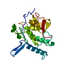
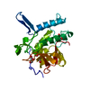
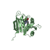
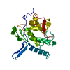
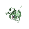
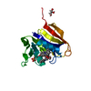
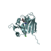
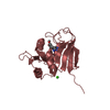
| Title | Structure of the Resuscitation Promoting Factor Interacting protein RipA mutated at C383 | ||||||
 Components Components | Peptidoglycan endopeptidase RipA | ||||||
 Keywords Keywords |  HYDROLASE / alpha-beta / cell wall hydrolase HYDROLASE / alpha-beta / cell wall hydrolase | ||||||
| Function / homology |  Function and homology information Function and homology informationcell wall organization or biogenesis / N-acetylmuramoyl-L-alanine amidase activity / Hydrolases; Acting on peptide bonds (peptidases) / cysteine-type peptidase activity / peptidoglycan-based cell wall / cell wall organization / proteolysis / extracellular regionSimilarity search - Function | ||||||
| Biological species |   Mycobacterium tuberculosis (bacteria) Mycobacterium tuberculosis (bacteria) | ||||||
| Method | X-RAY DIFFRACTION / SYNCHROTRON / MOLECULAR REPLACEMENT / Resolution: 0.97 Å | ||||||
 Authors Authors | Berisio, R. | ||||||
 Citation Citation | Journal: Acta Crystallogr.,Sect.D / Year: 2014 Title: Mutational and structural study of RipA, a key enzyme in Mycobacterium tuberculosis cell division: evidence for the L-to-D inversion of configuration of the catalytic cysteine. Authors: Squeglia, F. / Ruggiero, A. / Romano, M. / Vitagliano, L. / Berisio, R. | ||||||
| History |
|
- Structure visualization
| Structure viewer | Molecule: MolmilJmol/JSmol |
|---|
- Downloads & links
-Download
| PDBx/mmCIF format | 4q4g.cif.gz | 110.3 KB | Display | PDBx/mmCIF format |
|---|---|---|---|---|
| PDB format | pdb4q4g.ent.gz | 81.2 KB | Display | PDB format |
| PDBx/mmJSON format | 4q4g.json.gz | Tree view | PDBx/mmJSON format | |
| Others |  Other downloads Other downloads |
-Validation report
| Arichive directory | https://data.pdbj.org/pub/pdb/validation_reports/q4/4q4gftp://data.pdbj.org/pub/pdb/validation_reports/q4/4q4g | HTTPS FTP |
|---|
-Related structure data


-Links
 PDBj
PDBj- Assembly
| Deposited unit | 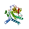
| ||||||||
|---|---|---|---|---|---|---|---|---|---|
| 1 |
| ||||||||
| Unit cell |
|
-Components
| #1: Protein | Mass: 49828.801 Da / Num. of mol.: 1 / Mutation: C383A Source method: isolated from a genetically manipulated source Source: (gene. exp.) Mycobacterium tuberculosis (bacteria) / Gene: ripA, Rv1477 / Production host: Escherichia coli (E. coli)References: UniProt: O53168, Hydrolases; Acting on peptide bonds (peptidases) |
|---|---|
| #2: Water | ChemComp-HOH / Water Mass: 18.015 Da / Num. of mol.: 395 / Source method: isolated from a natural source / Formula: H2O Mass: 18.015 Da / Num. of mol.: 395 / Source method: isolated from a natural source / Formula: H2O |
-Experimental details
-Experiment
| Experiment | Method: X-RAY DIFFRACTION / Number of used crystals: 1 |
|---|
- Sample preparation
| Crystal grow | Temperature: 293 K / Method: vapor diffusion, hanging drop / pH: 5.6 Details: 5 and 8 mg mL-1 protein concentration, respectively, and 8% (v/v) 2-Propanol, 16% (w/v) PEG4000 in 60 mM Sodium citrate trihydrate buffer, pH 5.6, VAPOR DIFFUSION, HANGING DROP, temperature 293K |
|---|
-Data collection
| Diffraction | Mean temperature: 100 K |
|---|---|
| Diffraction source | Source: SYNCHROTRON / Site: ESRF  / Beamline: BM14 / Wavelength: 0.9343 Å / Beamline: BM14 / Wavelength: 0.9343 Å |
| Detector | Type: MARMOSAIC 225 mm CCD / Detector: CCD / Date: Mar 3, 2011 |
| Radiation | Protocol: SINGLE WAVELENGTH / Monochromatic (M) / Laue (L): M / Scattering type: x-ray |
| Radiation wavelength | Wavelength: 0.9343 Å / Relative weight: 1 |
| Reflection | Resolution: 0.97→30 Å / Num. all: 97797 / Num. obs: 97211 / Observed criterion σ(F): 0 / Observed criterion σ(I): 0 |
| Reflection shell | Resolution: 0.97→1 Å / % possible all: 99.9 |
- Processing
| Software | Name: REFMAC / Version: 5.5.0110 / Classification: refinement | ||||||||||||||||||||||||||||||||||||||||||||||||||||||||||||||||||||||||||||||||||||||||||||||||||||
|---|---|---|---|---|---|---|---|---|---|---|---|---|---|---|---|---|---|---|---|---|---|---|---|---|---|---|---|---|---|---|---|---|---|---|---|---|---|---|---|---|---|---|---|---|---|---|---|---|---|---|---|---|---|---|---|---|---|---|---|---|---|---|---|---|---|---|---|---|---|---|---|---|---|---|---|---|---|---|---|---|---|---|---|---|---|---|---|---|---|---|---|---|---|---|---|---|---|---|---|---|---|
| Refinement | Method to determine structure: MOLECULAR REPLACEMENT / Resolution: 0.97→10.54 Å / Cor.coef. Fo:Fc: 0.981 / SU B: 0.382 / SU ML: 0.01 / ESU R: 0.02 / Stereochemistry target values: MAXIMUM LIKELIHOOD / Details: HYDROGENS HAVE BEEN ADDED IN THE RIDING POSITIONS
| ||||||||||||||||||||||||||||||||||||||||||||||||||||||||||||||||||||||||||||||||||||||||||||||||||||
| Solvent computation | Ion probe radii: 0.8 Å / Shrinkage radii: 0.8 Å / VDW probe radii: 1.4 Å / Solvent model: MASK | ||||||||||||||||||||||||||||||||||||||||||||||||||||||||||||||||||||||||||||||||||||||||||||||||||||
| Displacement parameters | Biso mean: 7.752 Å2
| ||||||||||||||||||||||||||||||||||||||||||||||||||||||||||||||||||||||||||||||||||||||||||||||||||||
| Refinement step | Cycle: LAST / Resolution: 0.97→10.54 Å
| ||||||||||||||||||||||||||||||||||||||||||||||||||||||||||||||||||||||||||||||||||||||||||||||||||||
| Refine LS restraints |
| ||||||||||||||||||||||||||||||||||||||||||||||||||||||||||||||||||||||||||||||||||||||||||||||||||||
| LS refinement shell | Resolution: 0.97→0.995 Å / Total num. of bins used: 20
|
