 Movie
Movie Controller

[English] 日本語
 Yorodumi
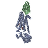
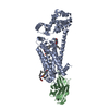
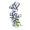
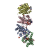
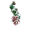
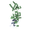
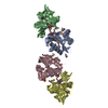
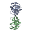
- PDB-4ldl: Structure of beta2 adrenoceptor bound to hydroxybenzylisoproteren... -
+ Open data

- Basic information
| Entry | Database: PDB / ID: 4ldl | ||||||
|---|---|---|---|---|---|---|---|
| Title | Structure of beta2 adrenoceptor bound to hydroxybenzylisoproterenol and an engineered nanobody | ||||||
 Components Components |
| ||||||
 Keywords Keywords | MEMBRANE PROTEIN/HYDROLASE / G protein coupled receptor / MEMBRANE PROTEIN-HYDROLASE complex | ||||||
| Function / homology |  Function and homology information Function and homology informationpositive regulation of mini excitatory postsynaptic potential / beta2-adrenergic receptor activity / negative regulation of smooth muscle contraction / AMPA selective glutamate receptor signaling pathway / norepinephrine-epinephrine-mediated vasodilation involved in regulation of systemic arterial blood pressure / positive regulation of autophagosome maturation / heat generation / norepinephrine binding / Adrenoceptors / positive regulation of lipophagy ...positive regulation of mini excitatory postsynaptic potential / beta2-adrenergic receptor activity / negative regulation of smooth muscle contraction / AMPA selective glutamate receptor signaling pathway / norepinephrine-epinephrine-mediated vasodilation involved in regulation of systemic arterial blood pressure / positive regulation of autophagosome maturation / heat generation / norepinephrine binding / Adrenoceptors / positive regulation of lipophagy / negative regulation of G protein-coupled receptor signaling pathway / negative regulation of multicellular organism growth / response to psychosocial stress / adrenergic receptor signaling pathway / endosome to lysosome transport / diet induced thermogenesis / positive regulation of cAMP/PKA signal transduction / adenylate cyclase binding / smooth muscle contraction / bone resorption / potassium channel regulator activity / positive regulation of bone mineralization / neuronal dense core vesicle / viral release from host cell by cytolysis / intercellular bridge / regulation of sodium ion transport / adenylate cyclase-activating adrenergic receptor signaling pathway / peptidoglycan catabolic process / brown fat cell differentiation / receptor-mediated endocytosis / response to cold / clathrin-coated endocytic vesicle membrane / adenylate cyclase-modulating G protein-coupled receptor signaling pathway / cellular response to amyloid-beta / cell wall macromolecule catabolic process / lysozyme / lysozyme activity / mitotic spindle / Cargo recognition for clathrin-mediated endocytosis / positive regulation of cold-induced thermogenesis / amyloid-beta binding / Clathrin-mediated endocytosis / microtubule cytoskeleton / G alpha (s) signalling events / transcription by RNA polymerase II / host cell cytoplasm / early endosome / cell surface receptor signaling pathway / lysosome / receptor complex / positive regulation of MAPK cascade / apical plasma membrane / endosome / endosome membrane / defense response to bacterium / Ub-specific processing proteases / cilium / ciliary basal body / protein-containing complex binding / Golgi apparatus / protein homodimerization activity / positive regulation of transcription by RNA polymerase II / identical protein binding / membrane / nucleus / plasma membrane Similarity search - Function | ||||||
| Biological species |  Enterobacteria phage T4 (virus) Enterobacteria phage T4 (virus) Homo sapiens (human) Homo sapiens (human) | ||||||
| Method |  X-RAY DIFFRACTION / SYNCHROTRON / MOLECULAR REPLACEMENT / Resolution: 3.1 Å X-RAY DIFFRACTION / SYNCHROTRON / MOLECULAR REPLACEMENT / Resolution: 3.1 Å | ||||||
 Authors Authors | Ring, A.M. / Manglik, A. / Kruse, A.C. / Enos, M.D. / Weis, W.I. / Garcia, K.C. / Kobilka, B.K. | ||||||
 Citation Citation | Journal: Nature / Year: 2013 Title: Adrenaline-activated structure of beta 2-adrenoceptor stabilized by an engineered nanobody. Authors: Ring, A.M. / Manglik, A. / Kruse, A.C. / Enos, M.D. / Weis, W.I. / Garcia, K.C. / Kobilka, B.K. | ||||||
| History |
| ||||||
| Remark 650 | HELIX DETERMINATION METHOD: AUTHOR DETERMINED |
- Structure visualization
| Structure viewer | Molecule: MolmilJmol/JSmol |
|---|
- Downloads & links
-Download
| PDBx/mmCIF format | 4ldl.cif.gz | 245.8 KB | Display | PDBx/mmCIF format |
|---|---|---|---|---|
| PDB format | pdb4ldl.ent.gz | 197.3 KB | Display | PDB format |
| PDBx/mmJSON format | 4ldl.json.gz | Tree view | PDBx/mmJSON format | |
| Others |  Other downloads Other downloads |
-Validation report
| Arichive directory | https://data.pdbj.org/pub/pdb/validation_reports/ld/4ldlftp://data.pdbj.org/pub/pdb/validation_reports/ld/4ldl | HTTPS FTP |
|---|
-Related structure data
| Related structure data | 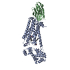 4ldeSC  4ldoC S: Starting model for refinement C: citing same article ( |
|---|---|
| Similar structure data |


-Links
 PDBj
PDBj


- Assembly
| Deposited unit | 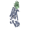
| ||||||||
|---|---|---|---|---|---|---|---|---|---|
| 1 |
| ||||||||
| Unit cell |
|
-Components
-Protein / Antibody , 2 types, 2 molecules AB
| #1: Protein | Mass: 53471.039 Da / Num. of mol.: 1 / Mutation: C918T, C962A, M1096T, M1098T, N1157E, C1265A Source method: isolated from a genetically manipulated source Source: (gene. exp.) Enterobacteria phage T4 (virus), (gene. exp.) Homo sapiens (human)Gene: ADRB2 / Plasmid: pVl1392 / Production host:   Spodoptera frugiperda (fall armyworm) / Strain (production host): Sf9 / References: UniProt: P00720, UniProt: P07550, lysozyme Spodoptera frugiperda (fall armyworm) / Strain (production host): Sf9 / References: UniProt: P00720, UniProt: P07550, lysozyme |
|---|---|
| #2: Antibody | Mass: 12949.375 Da / Num. of mol.: 1 Source method: isolated from a genetically manipulated source Details: Engineered fragment / Source: (gene. exp.)  |
-Non-polymers , 4 types, 5 molecules 


| #3: Chemical | ChemComp-XQC /  Mass: 317.380 Da / Num. of mol.: 1 / Source method: obtained synthetically / Formula: C18H23NO4 Mass: 317.380 Da / Num. of mol.: 1 / Source method: obtained synthetically / Formula: C18H23NO4 |
|---|---|
| #4: Chemical | ChemComp-NA /  Mass: 22.990 Da / Num. of mol.: 1 / Source method: obtained synthetically / Formula: Na Mass: 22.990 Da / Num. of mol.: 1 / Source method: obtained synthetically / Formula: Na |
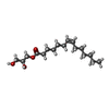
| #5: Chemical | ChemComp-1WV / ( Mass: 300.434 Da / Num. of mol.: 1 / Source method: obtained synthetically / Formula: C17H32O4 Mass: 300.434 Da / Num. of mol.: 1 / Source method: obtained synthetically / Formula: C17H32O4 |
| #6: Water | ChemComp-HOH / Mass: 18.015 Da / Num. of mol.: 2 / Source method: isolated from a natural source / Formula: H2O |
-Details
| Has protein modification | Y |
|---|
-Experimental details
-Experiment
| Experiment | Method: X-RAY DIFFRACTION / Number of used crystals: 9 |
|---|
- Sample preparation
| Crystal | Density Matthews: 3.77 Å3/Da / Density % sol: 67.35 % |
|---|---|
| Crystal grow | Temperature: 293 K / Method: lipidic cubic phase / pH: 6.5 Details: 100 mM MES pH 6.2-6.7, 40-100 mM ammonium phosphate dibasic, 18-24% PEG400, LIPIDIC CUBIC PHASE, temperature 293K |
-Data collection
| Diffraction | Mean temperature: 78 K |
|---|---|
| Diffraction source | Source: SYNCHROTRON / Site: APS  / Beamline: 23-ID-B / Wavelength: 1.033 Å / Beamline: 23-ID-B / Wavelength: 1.033 Å |
| Detector | Type: MARMOSAIC 300 mm CCD / Detector: CCD / Date: Dec 13, 2012 |
| Radiation | Monochromator: Double crystal cryo-cooled Si(111) / Protocol: SINGLE WAVELENGTH / Monochromatic (M) / Laue (L): M / Scattering type: x-ray |
| Radiation wavelength | Wavelength: 1.033 Å / Relative weight: 1 |
| Reflection | Resolution: 3.1→33.3 Å / Num. obs: 18053 / % possible obs: 94.5 % / Observed criterion σ(F): -3 / Redundancy: 4.6 % / Rmerge(I) obs: 0.177 / Net I/σ(I): 6.2 |
| Reflection shell | Resolution: 3.1→3.2 Å / Redundancy: 3.3 % / Rmerge(I) obs: 0.659 / Mean I/σ(I) obs: 1.9 / Num. unique all: 1789 / % possible all: 95.4 |
- Processing
| Software |
| |||||||||||||||||||||||||||||||||||||||||||||||||||||||||||||||||||||||||||||||||||||||||||||||||||||||||||||||||||||||||||||||||||||||||||||||||||||||||||||||||||||||||||||||||||||||||||||||||||||||||||||||||||||||||||||||||
|---|---|---|---|---|---|---|---|---|---|---|---|---|---|---|---|---|---|---|---|---|---|---|---|---|---|---|---|---|---|---|---|---|---|---|---|---|---|---|---|---|---|---|---|---|---|---|---|---|---|---|---|---|---|---|---|---|---|---|---|---|---|---|---|---|---|---|---|---|---|---|---|---|---|---|---|---|---|---|---|---|---|---|---|---|---|---|---|---|---|---|---|---|---|---|---|---|---|---|---|---|---|---|---|---|---|---|---|---|---|---|---|---|---|---|---|---|---|---|---|---|---|---|---|---|---|---|---|---|---|---|---|---|---|---|---|---|---|---|---|---|---|---|---|---|---|---|---|---|---|---|---|---|---|---|---|---|---|---|---|---|---|---|---|---|---|---|---|---|---|---|---|---|---|---|---|---|---|---|---|---|---|---|---|---|---|---|---|---|---|---|---|---|---|---|---|---|---|---|---|---|---|---|---|---|---|---|---|---|---|---|---|---|---|---|---|---|---|---|---|---|---|---|---|---|---|---|
| Refinement | Method to determine structure: MOLECULAR REPLACEMENT Starting model: PDB ENTRY 4LDE Resolution: 3.1→33.265 Å / SU ML: 0.45 / Cross valid method: THROUGHOUT / σ(F): 1.38 / Phase error: 27.17 / Stereochemistry target values: ML
| |||||||||||||||||||||||||||||||||||||||||||||||||||||||||||||||||||||||||||||||||||||||||||||||||||||||||||||||||||||||||||||||||||||||||||||||||||||||||||||||||||||||||||||||||||||||||||||||||||||||||||||||||||||||||||||||||
| Solvent computation | Shrinkage radii: 0.9 Å / VDW probe radii: 1.11 Å / Solvent model: FLAT BULK SOLVENT MODEL | |||||||||||||||||||||||||||||||||||||||||||||||||||||||||||||||||||||||||||||||||||||||||||||||||||||||||||||||||||||||||||||||||||||||||||||||||||||||||||||||||||||||||||||||||||||||||||||||||||||||||||||||||||||||||||||||||
| Refinement step | Cycle: LAST / Resolution: 3.1→33.265 Å
| |||||||||||||||||||||||||||||||||||||||||||||||||||||||||||||||||||||||||||||||||||||||||||||||||||||||||||||||||||||||||||||||||||||||||||||||||||||||||||||||||||||||||||||||||||||||||||||||||||||||||||||||||||||||||||||||||
| Refine LS restraints |
| |||||||||||||||||||||||||||||||||||||||||||||||||||||||||||||||||||||||||||||||||||||||||||||||||||||||||||||||||||||||||||||||||||||||||||||||||||||||||||||||||||||||||||||||||||||||||||||||||||||||||||||||||||||||||||||||||
| LS refinement shell |
| |||||||||||||||||||||||||||||||||||||||||||||||||||||||||||||||||||||||||||||||||||||||||||||||||||||||||||||||||||||||||||||||||||||||||||||||||||||||||||||||||||||||||||||||||||||||||||||||||||||||||||||||||||||||||||||||||
| Refinement TLS params. | Method: refined / Refine-ID: X-RAY DIFFRACTION
| |||||||||||||||||||||||||||||||||||||||||||||||||||||||||||||||||||||||||||||||||||||||||||||||||||||||||||||||||||||||||||||||||||||||||||||||||||||||||||||||||||||||||||||||||||||||||||||||||||||||||||||||||||||||||||||||||
| Refinement TLS group |
|
