 Movie
Movie Controller

[English] 日本語
 Yorodumi
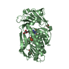
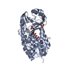
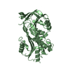
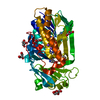
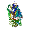
- PDB-3g8w: Crystal structure of a probable acetyltransferase from Staphyloco... -
+ Open data

- Basic information
| Entry | Database: PDB / ID: 3g8w | ||||||
|---|---|---|---|---|---|---|---|
| Title | Crystal structure of a probable acetyltransferase from Staphylococcus epidermidis ATCC 12228 | ||||||
 Components Components | Lactococcal prophage ps3 protein 05 | ||||||
 Keywords Keywords | TRANSFERASE / APC61042 / acetyltransferase / Staphylococcus epidermidis ATCC 12228 / structural genomics / PSI-2 / Protein Structure Initiative / Midwest Center for Structural Genomics / MCSG | ||||||
| Function / homology |  Function and homology information Function and homology informationN-acetyltransferase activity / acyltransferase activity, transferring groups other than amino-acyl groups Similarity search - Function | ||||||
| Biological species |  Staphylococcus epidermidis ATCC 12228 (bacteria) Staphylococcus epidermidis ATCC 12228 (bacteria) | ||||||
| Method |  X-RAY DIFFRACTION / SYNCHROTRON / MAD / Resolution: 2.7 Å X-RAY DIFFRACTION / SYNCHROTRON / MAD / Resolution: 2.7 Å | ||||||
 Authors Authors | Tan, K. / Sather, A. / Marshall, N. / Clancy, S. / Joachimiak, A. / Midwest Center for Structural Genomics (MCSG) | ||||||
 Citation Citation | Journal: To be Published Title: The crystal structure of a probable acetyltransferase from Staphylococcus epidermidis ATCC 12228. Authors: Tan, K. / Sather, A. / Marshall, N. / Clancy, S. / Joachimiak, A. | ||||||
| History |
|
- Structure visualization
| Structure viewer | Molecule: MolmilJmol/JSmol |
|---|
- Downloads & links
-Download
| PDBx/mmCIF format | 3g8w.cif.gz | 146.7 KB | Display | PDBx/mmCIF format |
|---|---|---|---|---|
| PDB format | pdb3g8w.ent.gz | 116.3 KB | Display | PDB format |
| PDBx/mmJSON format | 3g8w.json.gz | Tree view | PDBx/mmJSON format | |
| Others |  Other downloads Other downloads |
-Validation report
| Arichive directory | https://data.pdbj.org/pub/pdb/validation_reports/g8/3g8wftp://data.pdbj.org/pub/pdb/validation_reports/g8/3g8w | HTTPS FTP |
|---|
-Related structure data
| Similar structure data | |
|---|---|
| Other databases |


-Links
 PDBj
PDBj


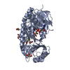
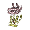
- Assembly
| Deposited unit | 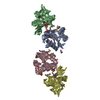
| ||||||||
|---|---|---|---|---|---|---|---|---|---|
| 1 | 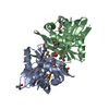
| ||||||||
| 2 | 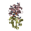
| ||||||||
| Unit cell |
| ||||||||
| Details | AUTHORS STATE THAT THE BIOLOGICAL UNIT IS EXPERIMENTALLY UNKNOWN. IT IS LIKELY A DIMER WITH THE ASSEMBLY SHOWN IN REMARK 350. |
-Components
| #1: Protein | Mass: 19975.586 Da / Num. of mol.: 4 Source method: isolated from a genetically manipulated source Source: (gene. exp.) Staphylococcus epidermidis ATCC 12228 (bacteria)Gene: SE_0588 / Plasmid: pMCSG19 / Production host: #2: Chemical | ChemComp-NHE /   Mass: 207.290 Da / Num. of mol.: 12 / Source method: obtained synthetically / Formula: C8H17NO3S / Comment: pH buffer*YM Mass: 207.290 Da / Num. of mol.: 12 / Source method: obtained synthetically / Formula: C8H17NO3S / Comment: pH buffer*YM#3: Chemical | ChemComp-FLC / |   Mass: 189.100 Da / Num. of mol.: 1 / Source method: obtained synthetically / Formula: C6H5O7 Mass: 189.100 Da / Num. of mol.: 1 / Source method: obtained synthetically / Formula: C6H5O7#4: Water | ChemComp-HOH / |  Mass: 18.015 Da / Num. of mol.: 21 / Source method: isolated from a natural source / Formula: H2O Mass: 18.015 Da / Num. of mol.: 21 / Source method: isolated from a natural source / Formula: H2OHas protein modification | Y | |
|---|
-Experimental details
-Experiment
| Experiment | Method: X-RAY DIFFRACTION / Number of used crystals: 1 |
|---|
- Sample preparation
| Crystal | Density Matthews: 3.69 Å3/Da / Density % sol: 66.69 % |
|---|---|
| Crystal grow | Temperature: 297 K / Method: vapor diffusion, sitting drop / pH: 9.5 Details: 0.1M CHES, 1.0M Sodium citrate, pH 9.5, VAPOR DIFFUSION, SITTING DROP, temperature 297K |
-Data collection
| Diffraction | Mean temperature: 100 K | |||||||||
|---|---|---|---|---|---|---|---|---|---|---|
| Diffraction source | Source: SYNCHROTRON / Site: APS  / Beamline: 19-ID / Wavelength: 0.97929, 0.97940 / Beamline: 19-ID / Wavelength: 0.97929, 0.97940 | |||||||||
| Detector | Type: ADSC QUANTUM 315 / Detector: CCD / Date: Nov 15, 2008 / Details: mirror | |||||||||
| Radiation | Monochromator: Si(111) crystal / Protocol: MAD / Monochromatic (M) / Laue (L): M / Scattering type: x-ray | |||||||||
| Radiation wavelength |
| |||||||||
| Reflection | Resolution: 2.7→50 Å / Num. all: 32550 / Num. obs: 32550 / % possible obs: 99.6 % / Observed criterion σ(F): 0 / Observed criterion σ(I): 0 / Redundancy: 6.5 % / Rmerge(I) obs: 0.093 / Net I/σ(I): 37.8 | |||||||||
| Reflection shell | Resolution: 2.7→2.75 Å / Redundancy: 6.8 % / Rmerge(I) obs: 0.759 / Mean I/σ(I) obs: 2.7 / Num. unique all: 1598 / % possible all: 100 |
- Processing
| Software |
| ||||||||||||||||||||||||||||||||||||||||||||||||||||||||||||||||||||||||||||||||||||||||||||||||||||||||||||||||||||||||||||||||||||||||||||||||||||||||||||||||||||||||||
|---|---|---|---|---|---|---|---|---|---|---|---|---|---|---|---|---|---|---|---|---|---|---|---|---|---|---|---|---|---|---|---|---|---|---|---|---|---|---|---|---|---|---|---|---|---|---|---|---|---|---|---|---|---|---|---|---|---|---|---|---|---|---|---|---|---|---|---|---|---|---|---|---|---|---|---|---|---|---|---|---|---|---|---|---|---|---|---|---|---|---|---|---|---|---|---|---|---|---|---|---|---|---|---|---|---|---|---|---|---|---|---|---|---|---|---|---|---|---|---|---|---|---|---|---|---|---|---|---|---|---|---|---|---|---|---|---|---|---|---|---|---|---|---|---|---|---|---|---|---|---|---|---|---|---|---|---|---|---|---|---|---|---|---|---|---|---|---|---|---|---|---|
| Refinement | Method to determine structure: MAD / Resolution: 2.7→49.39 Å / Cor.coef. Fo:Fc: 0.941 / Cor.coef. Fo:Fc free: 0.908 / SU B: 23.19 / SU ML: 0.223 / TLS residual ADP flag: LIKELY RESIDUAL / Cross valid method: THROUGHOUT / σ(F): 0 / σ(I): 0 / ESU R: 0.46 / ESU R Free: 0.298 / Stereochemistry target values: MAXIMUM LIKELIHOOD Details: HYDROGENS HAVE BEEN ADDED IN THE RIDING POSITIONS. Monomer D is partially disordered.
| ||||||||||||||||||||||||||||||||||||||||||||||||||||||||||||||||||||||||||||||||||||||||||||||||||||||||||||||||||||||||||||||||||||||||||||||||||||||||||||||||||||||||||
| Solvent computation | Ion probe radii: 0.8 Å / Shrinkage radii: 0.8 Å / VDW probe radii: 1.2 Å / Solvent model: MASK | ||||||||||||||||||||||||||||||||||||||||||||||||||||||||||||||||||||||||||||||||||||||||||||||||||||||||||||||||||||||||||||||||||||||||||||||||||||||||||||||||||||||||||
| Displacement parameters | Biso mean: 44.13 Å2
| ||||||||||||||||||||||||||||||||||||||||||||||||||||||||||||||||||||||||||||||||||||||||||||||||||||||||||||||||||||||||||||||||||||||||||||||||||||||||||||||||||||||||||
| Refinement step | Cycle: LAST / Resolution: 2.7→49.39 Å
| ||||||||||||||||||||||||||||||||||||||||||||||||||||||||||||||||||||||||||||||||||||||||||||||||||||||||||||||||||||||||||||||||||||||||||||||||||||||||||||||||||||||||||
| Refine LS restraints |
| ||||||||||||||||||||||||||||||||||||||||||||||||||||||||||||||||||||||||||||||||||||||||||||||||||||||||||||||||||||||||||||||||||||||||||||||||||||||||||||||||||||||||||
| LS refinement shell | Resolution: 2.7→2.77 Å / Total num. of bins used: 20
| ||||||||||||||||||||||||||||||||||||||||||||||||||||||||||||||||||||||||||||||||||||||||||||||||||||||||||||||||||||||||||||||||||||||||||||||||||||||||||||||||||||||||||
| Refinement TLS params. | Method: refined / Refine-ID: X-RAY DIFFRACTION
| ||||||||||||||||||||||||||||||||||||||||||||||||||||||||||||||||||||||||||||||||||||||||||||||||||||||||||||||||||||||||||||||||||||||||||||||||||||||||||||||||||||||||||
| Refinement TLS group |
|
