 Movie
Movie Controller


+ Open data

- Basic information
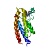
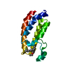
| Entry | Database: PDB / ID: 4lc2 | ||||||
|---|---|---|---|---|---|---|---|
| Title | Crystal structure of the bromodomain of human BRPF1B | ||||||
 Components Components | Peregrin | ||||||
 Keywords Keywords | DNA BINDING PROTEIN / Bromodomain and PHD finger-containing protein 1 / Protein Br140 / Structural Genomics Consortium / SGC | ||||||
| Function / homology |  Function and homology information Function and homology informationacetyltransferase activator activity / regulation of developmental process / MOZ/MORF histone acetyltransferase complex / regulation of hemopoiesis / histone acetyltransferase complex / Regulation of TP53 Activity through Acetylation / HATs acetylate histones / chromatin remodeling / regulation of transcription by RNA polymerase II / regulation of DNA-templated transcription ...acetyltransferase activator activity / regulation of developmental process / MOZ/MORF histone acetyltransferase complex / regulation of hemopoiesis / histone acetyltransferase complex / Regulation of TP53 Activity through Acetylation / HATs acetylate histones / chromatin remodeling / regulation of transcription by RNA polymerase II / regulation of DNA-templated transcription / positive regulation of DNA-templated transcription / DNA binding / zinc ion binding / nucleoplasm / nucleus / plasma membrane / cytoplasm / cytosol Similarity search - Function | ||||||
| Biological species |  Homo sapiens (human) Homo sapiens (human) | ||||||
| Method |  X-RAY DIFFRACTION / MOLECULAR REPLACEMENT / molecular replacement / Resolution: 1.65 Å X-RAY DIFFRACTION / MOLECULAR REPLACEMENT / molecular replacement / Resolution: 1.65 Å | ||||||
 Authors Authors | Tallant, C. / Nunez-Alonso, G. / Savitsky, P. / Picaud, S. / Filippakopoulos, P. / von Delft, F. / Arrowsmith, C.H. / Edwards, A.M. / Bountra, C. / Knapp, S. / Structural Genomics Consortium (SGC) | ||||||
 Citation Citation | Journal: TO BE PUBLISHED Title: Crystal structure of the bromodomain of human BRPF1B Authors: Tallant, C. / Nunez-Alonso, G. / Savitsky, P. / Picaud, S. / Filippakopoulos, P. / von Delft, F. / Arrowsmith, H.C. / Edwards, M.A. / Bountra, C. / Knapp, S. | ||||||
| History |
|
- Structure visualization
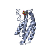
| Structure viewer | Molecule: MolmilJmol/JSmol |
|---|
- Downloads & links
-Download
| PDBx/mmCIF format | 4lc2.cif.gz | 64.6 KB | Display | PDBx/mmCIF format |
|---|---|---|---|---|
| PDB format | pdb4lc2.ent.gz | 47 KB | Display | PDB format |
| PDBx/mmJSON format | 4lc2.json.gz | Tree view | PDBx/mmJSON format | |
| Others |  Other downloads Other downloads |
-Validation report
| Arichive directory | https://data.pdbj.org/pub/pdb/validation_reports/lc/4lc2ftp://data.pdbj.org/pub/pdb/validation_reports/lc/4lc2 | HTTPS FTP |
|---|
-Related structure data
| Related structure data |  1dvvS  1x0jS 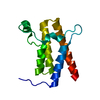 2grcS 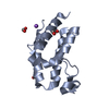 2oo1S 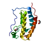 2ossS 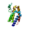 2ouoS 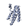 3d7cS 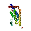 3daiS 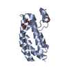 3dwyS 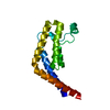 3hmhS S: Starting model for refinement |
|---|---|
| Similar structure data |


-Links
 PDBj
PDBj


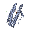
- Assembly
| Deposited unit | 
| |||||||||||||||
|---|---|---|---|---|---|---|---|---|---|---|---|---|---|---|---|---|
| 1 |
| |||||||||||||||
| Unit cell |
| |||||||||||||||
| Components on special symmetry positions |
|
-Components
| #1: Protein | Mass: 13703.698 Da / Num. of mol.: 1 / Fragment: unp residues 626-740 Source method: isolated from a genetically manipulated source Source: (gene. exp.) Homo sapiens (human) / Gene: BR140, BRPF1, BRPF1B / Plasmid: pNIC28-Bsa4 / Production host:  |
|---|---|
| #2: Chemical | ChemComp-NO3 /   Mass: 62.005 Da / Num. of mol.: 1 / Source method: obtained synthetically / Formula: NO3 Mass: 62.005 Da / Num. of mol.: 1 / Source method: obtained synthetically / Formula: NO3 |
| #3: Chemical | ChemComp-EDO /   Mass: 62.068 Da / Num. of mol.: 1 / Source method: obtained synthetically / Formula: C2H6O2 Mass: 62.068 Da / Num. of mol.: 1 / Source method: obtained synthetically / Formula: C2H6O2 |
| #4: Water | ChemComp-HOH /  Mass: 18.015 Da / Num. of mol.: 121 / Source method: isolated from a natural source / Formula: H2O Mass: 18.015 Da / Num. of mol.: 121 / Source method: isolated from a natural source / Formula: H2O |
-Experimental details
-Experiment
| Experiment | Method: X-RAY DIFFRACTION / Number of used crystals: 1 |
|---|
- Sample preparation
| Crystal | Density Matthews: 2.48 Å3/Da / Density % sol: 50.45 % |
|---|---|
| Crystal grow | Temperature: 277 K / Method: vapor diffusion, sitting drop / pH: 7.8 Details: 20% PEG3350 0.1 M bis tris propane, 10% ethylene glycol 0.15 M sodium nitrate, pH 7.8, VAPOR DIFFUSION, SITTING DROP, temperature 277K |
-Data collection
| Diffraction | Mean temperature: 100 K | |||||||||||||||||||||||||||||||||||||||||||||||||||||||||||||||||||||||||||||
|---|---|---|---|---|---|---|---|---|---|---|---|---|---|---|---|---|---|---|---|---|---|---|---|---|---|---|---|---|---|---|---|---|---|---|---|---|---|---|---|---|---|---|---|---|---|---|---|---|---|---|---|---|---|---|---|---|---|---|---|---|---|---|---|---|---|---|---|---|---|---|---|---|---|---|---|---|---|---|
| Diffraction source | Source: ROTATING ANODE / Type: RIGAKU FR-E SUPERBRIGHT / Wavelength: 1.5418 Å | |||||||||||||||||||||||||||||||||||||||||||||||||||||||||||||||||||||||||||||
| Detector | Type: RIGAKU RAXIS IV / Detector: IMAGE PLATE / Date: Jun 14, 2013 | |||||||||||||||||||||||||||||||||||||||||||||||||||||||||||||||||||||||||||||
| Radiation | Protocol: SINGLE WAVELENGTH / Monochromatic (M) / Laue (L): M / Scattering type: x-ray | |||||||||||||||||||||||||||||||||||||||||||||||||||||||||||||||||||||||||||||
| Radiation wavelength | Wavelength: 1.5418 Å / Relative weight: 1 | |||||||||||||||||||||||||||||||||||||||||||||||||||||||||||||||||||||||||||||
| Reflection | Resolution: 1.65→19 Å / Num. all: 16843 / Num. obs: 16793 / % possible obs: 99.7 % / Redundancy: 6.7 % / Biso Wilson estimate: 20.6 Å2 / Rmerge(I) obs: 0.026 / Rsym value: 0.026 / Net I/σ(I): 40.1 | |||||||||||||||||||||||||||||||||||||||||||||||||||||||||||||||||||||||||||||
| Reflection shell | Diffraction-ID: 1
|
-Phasing
| Phasing | Method: molecular replacement | |||||||||
|---|---|---|---|---|---|---|---|---|---|---|
| Phasing MR | Rfactor: 55.63 / Model details: Phaser MODE: MR_AUTO
|
- Processing
| Software |
| |||||||||||||||||||||||||||||||||||||||||||||||||||||||||||||||||||||||||||
|---|---|---|---|---|---|---|---|---|---|---|---|---|---|---|---|---|---|---|---|---|---|---|---|---|---|---|---|---|---|---|---|---|---|---|---|---|---|---|---|---|---|---|---|---|---|---|---|---|---|---|---|---|---|---|---|---|---|---|---|---|---|---|---|---|---|---|---|---|---|---|---|---|---|---|---|---|
| Refinement | Method to determine structure: MOLECULAR REPLACEMENT Starting model: Ensemble of PDB ENTRIES 1DVV, 1X0J, 3DAI, 3HMH, 2GRC, 2OO1, 2OSS, 2OUO, 3D7C, 3DWY Resolution: 1.65→19 Å / Cor.coef. Fo:Fc: 0.962 / Cor.coef. Fo:Fc free: 0.944 / WRfactor Rfree: 0.2241 / WRfactor Rwork: 0.1754 / Occupancy max: 1 / Occupancy min: 0.5 / FOM work R set: 0.8648 / SU B: 3.331 / SU ML: 0.059 / SU R Cruickshank DPI: 0.0901 / SU Rfree: 0.0967 / Cross valid method: THROUGHOUT / σ(F): 0 / ESU R: 0.09 / ESU R Free: 0.097 / Stereochemistry target values: MAXIMUM LIKELIHOOD Details: HYDROGENS HAVE BEEN ADDED IN THE RIDING POSITIONS U VALUES: WITH TLS ADDED
| |||||||||||||||||||||||||||||||||||||||||||||||||||||||||||||||||||||||||||
| Solvent computation | Ion probe radii: 0.8 Å / Shrinkage radii: 0.8 Å / VDW probe radii: 1.2 Å / Solvent model: MASK | |||||||||||||||||||||||||||||||||||||||||||||||||||||||||||||||||||||||||||
| Displacement parameters | Biso max: 119.16 Å2 / Biso mean: 25.5295 Å2 / Biso min: 8.91 Å2
| |||||||||||||||||||||||||||||||||||||||||||||||||||||||||||||||||||||||||||
| Refinement step | Cycle: LAST / Resolution: 1.65→19 Å
| |||||||||||||||||||||||||||||||||||||||||||||||||||||||||||||||||||||||||||
| Refine LS restraints |
| |||||||||||||||||||||||||||||||||||||||||||||||||||||||||||||||||||||||||||
| LS refinement shell | Resolution: 1.65→1.693 Å / Total num. of bins used: 20
| |||||||||||||||||||||||||||||||||||||||||||||||||||||||||||||||||||||||||||
| Refinement TLS params. | Method: refined / Origin x: 12.7879 Å / Origin y: 25.2852 Å / Origin z: 11.1287 Å
|
