 Movie
Movie Controller


+ Open data

- Basic information
| Entry | Database: PDB / ID: 4l3b | ||||||
|---|---|---|---|---|---|---|---|
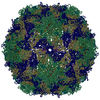
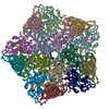
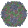
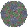
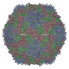
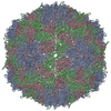
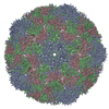
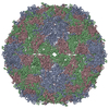
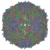
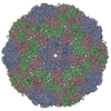
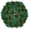
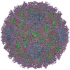
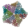
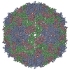
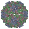
| Title | X-ray structure of the HRV2 A particle uncoating intermediate | ||||||
 Components Components |
| ||||||
 Keywords Keywords |  VIRUS / HRV2 capsid VIRUS / HRV2 capsid | ||||||
| Function / homology |  Function and homology information Function and homology informationsymbiont-mediated suppression of host cytoplasmic pattern recognition receptor signaling pathway via inhibition of RIG-I activity / picornain 2A / symbiont-mediated suppression of host mRNA export from nucleus / ribonucleoside triphosphate phosphatase activity / symbiont genome entry into host cell via pore formation in plasma membrane / picornain 3C / T=pseudo3 icosahedral viral capsid / host cell cytoplasmic vesicle membrane / endocytosis involved in viral entry into host cell / : ...symbiont-mediated suppression of host cytoplasmic pattern recognition receptor signaling pathway via inhibition of RIG-I activity / picornain 2A / symbiont-mediated suppression of host mRNA export from nucleus / ribonucleoside triphosphate phosphatase activity / symbiont genome entry into host cell via pore formation in plasma membrane / picornain 3C / T=pseudo3 icosahedral viral capsid / host cell cytoplasmic vesicle membrane / endocytosis involved in viral entry into host cell / : / nucleoside-triphosphate phosphatase / protein complex oligomerization / monoatomic ion channel activity / RNA helicase activity / DNA replication / induction by virus of host autophagy / RNA-directed RNA polymerase / symbiont-mediated suppression of host gene expression / viral RNA genome replication / cysteine-type endopeptidase activity / RNA-dependent RNA polymerase activity / DNA-templated transcription / host cell nucleus / virion attachment to host cell / structural molecule activity / proteolysis / RNA binding / ATP binding / membrane / metal ion bindingSimilarity search - Function | ||||||
| Biological species |  Human rhinovirus A2 Human rhinovirus A2 | ||||||
| Method | X-RAY DIFFRACTION / SYNCHROTRON / MOLECULAR REPLACEMENT / Resolution: 6.5 Å | ||||||
 Authors Authors | Vives-Adrian, L. / Querol-Audi, J. / Garriga, D. / Pous, J. / Verdaguer, N. | ||||||
 Citation Citation | Journal: Proc Natl Acad Sci U S A / Year: 2013 Title: Uncoating of common cold virus is preceded by RNA switching as determined by X-ray and cryo-EM analyses of the subviral A-particle. Authors: Angela Pickl-Herk / Daniel Luque / Laia Vives-Adrián / Jordi Querol-Audí / Damià Garriga / Benes L Trus / Nuria Verdaguer / Dieter Blaas / José R Castón /  Abstract: During infection, viruses undergo conformational changes that lead to delivery of their genome into host cytosol. In human rhinovirus A2, this conversion is triggered by exposure to acid pH in the ...During infection, viruses undergo conformational changes that lead to delivery of their genome into host cytosol. In human rhinovirus A2, this conversion is triggered by exposure to acid pH in the endosome. The first subviral intermediate, the A-particle, is expanded and has lost the internal viral protein 4 (VP4), but retains its RNA genome. The nucleic acid is subsequently released, presumably through one of the large pores that open at the icosahedral twofold axes, and is transferred along a conduit in the endosomal membrane; the remaining empty capsids, termed B-particles, are shuttled to lysosomes for degradation. Previous structural analyses revealed important differences between the native protein shell and the empty capsid. Nonetheless, little is known of A-particle architecture or conformation of the RNA core. Using 3D cryo-electron microscopy and X-ray crystallography, we found notable changes in RNA-protein contacts during conversion of native virus into the A-particle uncoating intermediate. In the native virion, we confirmed interaction of nucleotide(s) with Trp(38) of VP2 and identified additional contacts with the VP1 N terminus. Study of A-particle structure showed that the VP2 contact is maintained, that VP1 interactions are lost after exit of the VP1 N-terminal extension, and that the RNA also interacts with residues of the VP3 N terminus at the fivefold axis. These associations lead to formation of a well-ordered RNA layer beneath the protein shell, suggesting that these interactions guide ordered RNA egress. | ||||||
| History |
|
- Structure visualization
| Structure viewer | Molecule: MolmilJmol/JSmol |
|---|
- Downloads & links
-Download
| PDBx/mmCIF format | 4l3b.cif.gz | 153.8 KB | Display | PDBx/mmCIF format |
|---|---|---|---|---|
| PDB format | pdb4l3b.ent.gz | 120 KB | Display | PDB format |
| PDBx/mmJSON format | 4l3b.json.gz | Tree view | PDBx/mmJSON format | |
| Others |  Other downloads Other downloads |
-Validation report
| Arichive directory | https://data.pdbj.org/pub/pdb/validation_reports/l3/4l3bftp://data.pdbj.org/pub/pdb/validation_reports/l3/4l3b | HTTPS FTP |
|---|
-Related structure data
| Related structure data | 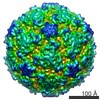 2106C 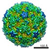 2107C 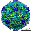 2108C 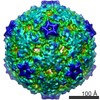 2109C  3tn9S S: Starting model for refinement C: citing same article ( |
|---|---|
| Similar structure data |

-Links
 PDBj
PDBj

- Assembly
| Deposited unit | 
| ||||||||||||||||||||||||||||||||||||||||||||||||
|---|---|---|---|---|---|---|---|---|---|---|---|---|---|---|---|---|---|---|---|---|---|---|---|---|---|---|---|---|---|---|---|---|---|---|---|---|---|---|---|---|---|---|---|---|---|---|---|---|---|
| 1 | x 60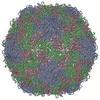
| ||||||||||||||||||||||||||||||||||||||||||||||||
| 2 |
| ||||||||||||||||||||||||||||||||||||||||||||||||
| 3 | x 5
| ||||||||||||||||||||||||||||||||||||||||||||||||
| 4 | x 6
| ||||||||||||||||||||||||||||||||||||||||||||||||
| 5 | 
| ||||||||||||||||||||||||||||||||||||||||||||||||
| 6 | x 15
| ||||||||||||||||||||||||||||||||||||||||||||||||
| Unit cell |
| ||||||||||||||||||||||||||||||||||||||||||||||||
| Symmetry | Point symmetry: (Schoenflies symbol: I (icosahedral)) | ||||||||||||||||||||||||||||||||||||||||||||||||
| Noncrystallographic symmetry (NCS) | NCS oper:
|
-Components
| #1: Protein | Mass: 32924.797 Da / Num. of mol.: 1 / Fragment: UNP residues 568-856 / Source method: isolated from a natural source / Source: (natural) Human rhinovirus A2 / References: UniProt: P04936 |
|---|---|
| #2: Protein | Mass: 29009.588 Da / Num. of mol.: 1 / Fragment: UNP residues 70-330 / Source method: isolated from a natural source / Source: (natural) Human rhinovirus A2 / References: UniProt: P04936 |
| #3: Protein | Mass: 26107.793 Da / Num. of mol.: 1 / Fragment: UNP residues 331-567 / Source method: isolated from a natural source / Source: (natural) Human rhinovirus A2 / References: UniProt: P04936 |
-Experimental details
-Experiment
| Experiment | Method: X-RAY DIFFRACTION / Number of used crystals: 1 |
|---|
- Sample preparation
| Crystal | Density Matthews: 4.09 Å3/Da / Density % sol: 69.89 % |
|---|---|
| Crystal grow | Temperature: 293 K / Method: vapor diffusion, hanging drop / pH: 7.5 Details: 0.5 M ammonium sulfate, 1 M sodium/potassium phosphate, 5% glycerol, pH 7.5, VAPOR DIFFUSION, HANGING DROP, temperature 293K |
-Data collection
| Diffraction | Mean temperature: 100 K |
|---|---|
| Diffraction source | Source: SYNCHROTRON / Site: SLS  / Beamline: X06SA / Wavelength: 1 Å / Beamline: X06SA / Wavelength: 1 Å |
| Detector | Type: PSI PILATUS 6M / Detector: PIXEL / Date: May 4, 2012 |
| Radiation | Monochromator: double crystal Si(111) / Protocol: SINGLE WAVELENGTH / Monochromatic (M) / Laue (L): M / Scattering type: x-ray |
| Radiation wavelength | Wavelength: 1 Å / Relative weight: 1 |
| Reflection | Resolution: 4.5→262.63 Å / Num. obs: 15750 / % possible obs: 12.5 % / Observed criterion σ(F): 2 / Observed criterion σ(I): 1 / Rmerge(I) obs: 0.163 / Rsym value: 0.163 / Net I/σ(I): 4.4 |
| Reflection shell | Resolution: 4.5→4.74 Å / % possible all: 12.5 |
- Processing
| Software |
| |||||||||||||||||||||
|---|---|---|---|---|---|---|---|---|---|---|---|---|---|---|---|---|---|---|---|---|---|---|
| Refinement | Method to determine structure: MOLECULAR REPLACEMENT Starting model: PDB ENTRY 3TN9 Resolution: 6.5→121.39 Å / σ(F): 2 / Stereochemistry target values: Engh & Huber
| |||||||||||||||||||||
| Refinement step | Cycle: LAST / Resolution: 6.5→121.39 Å
|
