 Movie
Movie Controller


+ Open data

- Basic information
| Entry | Database: PDB / ID: 4dbp | ||||||
|---|---|---|---|---|---|---|---|
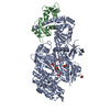
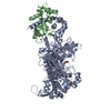
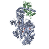
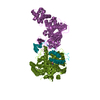
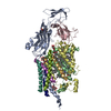
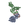
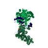
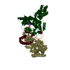
| Title | Myosin VI nucleotide-free (MDINSERT2) D179Y crystal structure | ||||||
 Components Components |
| ||||||
 Keywords Keywords | motor protein/Calcium binding protein / motor protein / motor protein-Calcium binding protein complex | ||||||
| Function / homology |  Function and homology information Function and homology informationnegative regulation of phospholipase C-activating phototransduction signaling pathway / myosin VI complex / myosin VI head/neck binding / myosin VII complex / photoreceptor cell axon guidance / negative regulation of opsin-mediated signaling pathway / rhabdomere development / rhabdomere / regulation of secretion / myosin V complex ...negative regulation of phospholipase C-activating phototransduction signaling pathway / myosin VI complex / myosin VI head/neck binding / myosin VII complex / photoreceptor cell axon guidance / negative regulation of opsin-mediated signaling pathway / rhabdomere development / rhabdomere / regulation of secretion / myosin V complex / detection of chemical stimulus involved in sensory perception of smell / kinetochore organization / autophagic cell death / G protein-coupled opsin signaling pathway / inner ear auditory receptor cell differentiation / actin filament-based movement / myosin V binding / channel regulator activity / myosin complex / clathrin-coated vesicle / inner ear morphogenesis / myosin heavy chain binding / muscle cell cellular homeostasis / microfilament motor activity / mitotic spindle pole / filamentous actin / microvillus / centriole replication / cytoskeletal motor activity / cellular response to ethanol / enzyme regulator activity / ruffle / clathrin-coated pit / centriole / actin filament organization / actin filament / filopodium / intracellular protein transport / DNA damage response, signal transduction by p53 class mediator / sensory perception of sound / ADP binding / microtubule cytoskeleton organization / spindle / ruffle membrane / endocytosis / actin filament binding / mitotic spindle / sensory perception of smell / intracellular protein localization / actin cytoskeleton / protein transport / cytoplasmic vesicle / midbody / cell cortex / nuclear membrane / calmodulin binding / calcium ion binding / centrosome / perinuclear region of cytoplasm / Golgi apparatus / nucleoplasm / ATP binding / metal ion binding / nucleus / plasma membrane / cytoplasm / cytosol Similarity search - Function | ||||||
| Biological species |   | ||||||
| Method |  X-RAY DIFFRACTION / SYNCHROTRON / MOLECULAR REPLACEMENT / Resolution: 2.2 Å X-RAY DIFFRACTION / SYNCHROTRON / MOLECULAR REPLACEMENT / Resolution: 2.2 Å | ||||||
 Authors Authors | Pylypenko, O. / Sweeney, H.L. / Houdusse, A. | ||||||
 Citation Citation | Journal: To be Published Title: Mutations in myosin VI that cause a loss of coordination between heads provide insights into the structural changes underlying force generation and the importance of gating Authors: Song, L. / Pylypenko, O. / Yang, Z. / Houdusse, A. / Sweeney, L.H. | ||||||
| History |
|
- Structure visualization
| Structure viewer | Molecule: MolmilJmol/JSmol |
|---|
- Downloads & links
-Download
| PDBx/mmCIF format | 4dbp.cif.gz | 406.2 KB | Display | PDBx/mmCIF format |
|---|---|---|---|---|
| PDB format | pdb4dbp.ent.gz | 326.4 KB | Display | PDB format |
| PDBx/mmJSON format | 4dbp.json.gz | Tree view | PDBx/mmJSON format | |
| Others |  Other downloads Other downloads |
-Validation report
| Arichive directory | https://data.pdbj.org/pub/pdb/validation_reports/db/4dbpftp://data.pdbj.org/pub/pdb/validation_reports/db/4dbp | HTTPS FTP |
|---|
-Related structure data
| Related structure data | 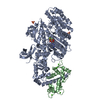 4dbqC 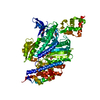 4dbrC  2bkhS S: Starting model for refinement C: citing same article ( |
|---|---|
| Similar structure data |


-Links
 PDBj
PDBj


- Assembly
| Deposited unit | 
| ||||||||
|---|---|---|---|---|---|---|---|---|---|
| 1 |
| ||||||||
| Unit cell |
|
-Components
-Protein , 2 types, 2 molecules AC
| #1: Protein | Mass: 93081.164 Da / Num. of mol.: 1 / Fragment: MOTOR DOMAIN-INSERT2, UNP residues 2-815 / Mutation: D179Y Source method: isolated from a genetically manipulated source Source: (gene. exp.)  Spodoptera frugiperda (fall armyworm) / References: UniProt: F1RQI7, UniProt: Q29122*PLUS Spodoptera frugiperda (fall armyworm) / References: UniProt: F1RQI7, UniProt: Q29122*PLUS |
|---|---|
| #2: Protein | Mass: 16825.520 Da / Num. of mol.: 1 Source method: isolated from a genetically manipulated source Source: (gene. exp.) Spodoptera frugiperda (fall armyworm) / References: UniProt: P62152 |
-Non-polymers , 5 types, 806 molecules 


| #3: Chemical |  Mass: 74.122 Da / Num. of mol.: 2 / Source method: obtained synthetically / Formula: C4H10O Mass: 74.122 Da / Num. of mol.: 2 / Source method: obtained synthetically / Formula: C4H10O#4: Chemical | ChemComp-EDO /  Mass: 62.068 Da / Num. of mol.: 18 / Source method: obtained synthetically / Formula: C2H6O2 Mass: 62.068 Da / Num. of mol.: 18 / Source method: obtained synthetically / Formula: C2H6O2#5: Chemical | ChemComp-IPA / |  Mass: 60.095 Da / Num. of mol.: 1 / Source method: obtained synthetically / Formula: C3H8O / Comment: alkaloid*YM Mass: 60.095 Da / Num. of mol.: 1 / Source method: obtained synthetically / Formula: C3H8O / Comment: alkaloid*YM#6: Chemical | ChemComp-CA /  Mass: 40.078 Da / Num. of mol.: 4 / Source method: obtained synthetically / Formula: Ca Mass: 40.078 Da / Num. of mol.: 4 / Source method: obtained synthetically / Formula: Ca#7: Water | ChemComp-HOH / | Mass: 18.015 Da / Num. of mol.: 781 / Source method: isolated from a natural source / Formula: H2O |
|---|
-Experimental details
-Experiment
| Experiment | Method: X-RAY DIFFRACTION / Number of used crystals: 1 |
|---|
- Sample preparation
| Crystal | Density Matthews: 2.93 Å3/Da / Density % sol: 58.05 % |
|---|---|
| Crystal grow | Temperature: 277 K / Method: vapor diffusion, hanging drop / pH: 7 Details: 4.5% P8K, 50 mM MOPS pH7,3% I-prop, 3% tert-but,1mM TCEP, pH 7.0, VAPOR DIFFUSION, HANGING DROP, temperature 277K |
-Data collection
| Diffraction | Mean temperature: 100 K |
|---|---|
| Diffraction source | Source: SYNCHROTRON / Site: ESRF  / Beamline: ID23-1 / Wavelength: 0.97625 Å / Beamline: ID23-1 / Wavelength: 0.97625 Å |
| Detector | Type: ADSC QUANTUM 315r / Detector: CCD / Date: Jun 15, 2008 |
| Radiation | Monochromator: Si(111) / Protocol: SINGLE WAVELENGTH / Monochromatic (M) / Laue (L): M / Scattering type: x-ray |
| Radiation wavelength | Wavelength: 0.97625 Å / Relative weight: 1 |
| Reflection | Resolution: 2.2→19.9 Å / Num. all: 64374 / Num. obs: 63904 / % possible obs: 99.3 % / Observed criterion σ(F): -3 / Observed criterion σ(I): -3 |
| Reflection shell | Resolution: 2.2→2.25 Å / % possible all: 98.8 |
- Processing
| Software |
| |||||||||||||||||||||||||||||||||||||||||||||||||||||||||||||||||||||||||||||||||||||||||||||||||||||||||||||||||||||||||||||
|---|---|---|---|---|---|---|---|---|---|---|---|---|---|---|---|---|---|---|---|---|---|---|---|---|---|---|---|---|---|---|---|---|---|---|---|---|---|---|---|---|---|---|---|---|---|---|---|---|---|---|---|---|---|---|---|---|---|---|---|---|---|---|---|---|---|---|---|---|---|---|---|---|---|---|---|---|---|---|---|---|---|---|---|---|---|---|---|---|---|---|---|---|---|---|---|---|---|---|---|---|---|---|---|---|---|---|---|---|---|---|---|---|---|---|---|---|---|---|---|---|---|---|---|---|---|---|
| Refinement | Method to determine structure: MOLECULAR REPLACEMENT Starting model: PDB entry 2BKH Resolution: 2.2→19.885 Å / SU ML: 0.65 / σ(F): 0 / Phase error: 21.01 / Stereochemistry target values: ML
| |||||||||||||||||||||||||||||||||||||||||||||||||||||||||||||||||||||||||||||||||||||||||||||||||||||||||||||||||||||||||||||
| Solvent computation | Shrinkage radii: 0.98 Å / VDW probe radii: 1.2 Å / Solvent model: FLAT BULK SOLVENT MODEL / Bsol: 40.641 Å2 / ksol: 0.314 e/Å3 | |||||||||||||||||||||||||||||||||||||||||||||||||||||||||||||||||||||||||||||||||||||||||||||||||||||||||||||||||||||||||||||
| Refinement step | Cycle: LAST / Resolution: 2.2→19.885 Å
| |||||||||||||||||||||||||||||||||||||||||||||||||||||||||||||||||||||||||||||||||||||||||||||||||||||||||||||||||||||||||||||
| Refine LS restraints |
| |||||||||||||||||||||||||||||||||||||||||||||||||||||||||||||||||||||||||||||||||||||||||||||||||||||||||||||||||||||||||||||
| LS refinement shell |
| |||||||||||||||||||||||||||||||||||||||||||||||||||||||||||||||||||||||||||||||||||||||||||||||||||||||||||||||||||||||||||||
| Refinement TLS params. | Method: refined / Refine-ID: X-RAY DIFFRACTION
| |||||||||||||||||||||||||||||||||||||||||||||||||||||||||||||||||||||||||||||||||||||||||||||||||||||||||||||||||||||||||||||
| Refinement TLS group |
|
