| 登録情報 | データベース: PDB / ID: 4awx
|
|---|
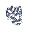
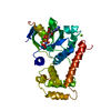
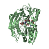
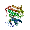
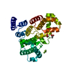
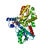
| タイトル | Moonlighting functions of FeoC in the regulation of ferrous iron transport in Feo |
|---|
 要素 要素 | - FERROUS IRON TRANSPORT PROTEIN B
- FERROUS IRON TRANSPORT PROTEIN C
|
|---|
 キーワード キーワード | METAL TRANSPORT |
|---|
| 機能・相同性 |  機能・相同性情報 機能・相同性情報
iron-sulfur cluster binding / iron ion binding / DNA binding類似検索 - 分子機能 Probable [Fe-S]-dependent transcriptional repressor / Transcriptional regulator HTH-type, FeoC / FeoC like transcriptional regulator / Helix Hairpins - #1770 / Winged helix-like DNA-binding domain superfamily/Winged helix DNA-binding domain / Helix Hairpins / Arc Repressor Mutant, subunit A / Winged helix DNA-binding domain superfamily / P-loop containing nucleotide triphosphate hydrolases / Winged helix-like DNA-binding domain superfamily ...Probable [Fe-S]-dependent transcriptional repressor / Transcriptional regulator HTH-type, FeoC / FeoC like transcriptional regulator / Helix Hairpins - #1770 / Winged helix-like DNA-binding domain superfamily/Winged helix DNA-binding domain / Helix Hairpins / Arc Repressor Mutant, subunit A / Winged helix DNA-binding domain superfamily / P-loop containing nucleotide triphosphate hydrolases / Winged helix-like DNA-binding domain superfamily / Rossmann fold / Orthogonal Bundle / 3-Layer(aba) Sandwich / Mainly Alpha / Alpha Beta類似検索 - ドメイン・相同性 NICKEL (II) ION / Probable [Fe-S]-dependent transcriptional repressor / : 類似検索 - 構成要素 |
|---|
| 生物種 |  KLEBSIELLA PNEUMONIAE (肺炎桿菌) KLEBSIELLA PNEUMONIAE (肺炎桿菌) |
|---|
| 手法 |  X線回折 / シンクロトロン / 分子置換 / 解像度: 2.3 Å X線回折 / シンクロトロン / 分子置換 / 解像度: 2.3 Å |
|---|
 データ登録者 データ登録者 | Hung, K.-W. / Tsai, J.-Y. / Juan, T.-H. / Hsu, Y.-L. / Hsiao, C.D. / Huang, T.-H. |
|---|
 引用 引用 | ジャーナル: J.Bacteriol. / 年: 2012
タイトル: Crystal Structure of the Klebsiella Pneumoniae Nfeob/Feoc Complex and Roles of Feoc in Regulation of Fe2+ Transport by the Bacterial Feo System.
著者: Hung, K.-W. / Tsai, J.-Y. / Juan, T.-H. / Hsiao, C.D. / Huang, T.-H. |
|---|
| 履歴 | | 登録 | 2012年6月6日 | 登録サイト: PDBE / 処理サイト: PDBE |
|---|
| 改定 1.0 | 2013年3月6日 | Provider: repository / タイプ: Initial release |
|---|
| 改定 1.1 | 2023年12月20日 | Group: Data collection / Database references ...Data collection / Database references / Derived calculations / Other / Refinement description
カテゴリ: chem_comp_atom / chem_comp_bond ...chem_comp_atom / chem_comp_bond / database_2 / pdbx_database_status / pdbx_initial_refinement_model / pdbx_struct_conn_angle / struct_conn / struct_site
Item: _database_2.pdbx_DOI / _database_2.pdbx_database_accession ..._database_2.pdbx_DOI / _database_2.pdbx_database_accession / _pdbx_database_status.status_code_sf / _pdbx_struct_conn_angle.ptnr1_auth_comp_id / _pdbx_struct_conn_angle.ptnr1_auth_seq_id / _pdbx_struct_conn_angle.ptnr1_label_asym_id / _pdbx_struct_conn_angle.ptnr1_label_atom_id / _pdbx_struct_conn_angle.ptnr1_label_comp_id / _pdbx_struct_conn_angle.ptnr1_label_seq_id / _pdbx_struct_conn_angle.ptnr1_symmetry / _pdbx_struct_conn_angle.ptnr2_auth_seq_id / _pdbx_struct_conn_angle.ptnr2_label_asym_id / _pdbx_struct_conn_angle.ptnr3_auth_comp_id / _pdbx_struct_conn_angle.ptnr3_auth_seq_id / _pdbx_struct_conn_angle.ptnr3_label_asym_id / _pdbx_struct_conn_angle.ptnr3_label_atom_id / _pdbx_struct_conn_angle.ptnr3_label_comp_id / _pdbx_struct_conn_angle.ptnr3_label_seq_id / _pdbx_struct_conn_angle.ptnr3_symmetry / _pdbx_struct_conn_angle.value / _struct_conn.pdbx_dist_value / _struct_conn.ptnr1_auth_comp_id / _struct_conn.ptnr1_auth_seq_id / _struct_conn.ptnr1_label_asym_id / _struct_conn.ptnr1_label_atom_id / _struct_conn.ptnr1_label_comp_id / _struct_conn.ptnr1_label_seq_id / _struct_conn.ptnr1_symmetry / _struct_conn.ptnr2_auth_comp_id / _struct_conn.ptnr2_auth_seq_id / _struct_conn.ptnr2_label_asym_id / _struct_conn.ptnr2_label_atom_id / _struct_conn.ptnr2_label_comp_id / _struct_conn.ptnr2_label_seq_id / _struct_conn.ptnr2_symmetry / _struct_site.pdbx_auth_asym_id / _struct_site.pdbx_auth_comp_id / _struct_site.pdbx_auth_seq_id |
|---|
|
|---|
 ムービー
ムービー コントローラー


 データを開く
 基本情報
基本情報 構造の表示
構造の表示 ダウンロードとリンク
 その他のダウンロード
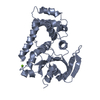


 PDBj
PDBj 集合体
集合体

 分子量: 96.063 Da / 分子数: 1 / 由来タイプ: 合成 / 式: SO4
 分子量: 58.693 Da / 分子数: 3 / 由来タイプ: 合成 / 式: Ni
 分子量: 18.015 Da / 分子数: 132 / 由来タイプ: 天然 / 式: H2O
 試料調製
試料調製 / ビームライン: BL13C1 / 波長: 0.976
  解析
解析