Mass: 18.015 Da / Num. of mol.: 298 / Source method: isolated from a natural source / Formula: H2O
Compound details
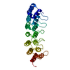
ENGINEERED RESIDUE IN CHAIN A, SER 711 TO ASP ENGINEERED RESIDUE IN CHAIN A, LYS 712 TO ALA ...ENGINEERED RESIDUE IN CHAIN A, SER 711 TO ASP ENGINEERED RESIDUE IN CHAIN A, LYS 712 TO ALA ENGINEERED RESIDUE IN CHAIN A, ASN 713 TO LEU ENGINEERED RESIDUE IN CHAIN A, TYR 800 TO PHE ENGINEERED RESIDUE IN CHAIN A, HIS 802 TO TRP ENGINEERED RESIDUE IN CHAIN A, LYS 803 TO ASN ENGINEERED RESIDUE IN CHAIN A, SER 805 TO TYR ENGINEERED RESIDUE IN CHAIN A, ALA 806 TO LYS ENGINEERED RESIDUE IN CHAIN A, MET 807 TO VAL ENGINEERED RESIDUE IN CHAIN A, ASP 809 TO SER ENGINEERED RESIDUE IN CHAIN A, LEU 810 TO GLU ENGINEERED RESIDUE IN CHAIN B, SER 711 TO ASP ENGINEERED RESIDUE IN CHAIN B, LYS 712 TO ALA ENGINEERED RESIDUE IN CHAIN B, ASN 713 TO LEU ENGINEERED RESIDUE IN CHAIN B, TYR 800 TO PHE ENGINEERED RESIDUE IN CHAIN B, HIS 802 TO TRP ENGINEERED RESIDUE IN CHAIN B, LYS 803 TO ASN ENGINEERED RESIDUE IN CHAIN B, SER 805 TO TYR ENGINEERED RESIDUE IN CHAIN B, ALA 806 TO LYS ENGINEERED RESIDUE IN CHAIN B, MET 807 TO VAL ENGINEERED RESIDUE IN CHAIN B, ASP 809 TO SER ENGINEERED RESIDUE IN CHAIN B, LEU 810 TO GLU
-
Experimental details
-
Experiment
Experiment
Method: X-RAY DIFFRACTION / Number of used crystals: 1
-
Sample preparation
Crystal
Density Matthews: 2.2 Å3/Da / Density % sol: 43.5 % / Description: NONE
Crystal grow
pH: 5 Details: PROTEIN SOLUTION: 20MM MES PH 6M 150MM NACL WELL SOLUTION: 0.1M NA ACETATE PH 5, 20% (W/V) PEG 8K, 6% (V/V) 2-PROPANOL CRYOPROTECTANT: WELL SOLUTION PLUS 15% (V/V) ETHYLENE GLYCOL
Resolution: 1.09→23.135 Å / SU ML: 0.24 / σ(F): 0.27 / Phase error: 17.48 / Stereochemistry target values: ML Details: RESIDUES -1 TO 0 AND 23-26 ARE DISORDERED IN CHAINS A AND B
Rfactor
Num. reflection
% reflection
Rfree
0.1806
4502
5 %
Rwork
0.158
-
-
obs
0.1592
90295
92.33 %
Solvent computation
Shrinkage radii: 0.77 Å / VDW probe radii: 0.9 Å / Solvent model: FLAT BULK SOLVENT MODEL / Bsol: 46.288 Å2 / ksol: 0.406 e/Å3
Displacement parameters
Biso mean: 15.8 Å2
Baniso -1
Baniso -2
Baniso -3
1-
0.4479 Å2
0 Å2
-0.0563 Å2
2-
-
-0.9722 Å2
0 Å2
3-
-
-
0.5243 Å2
Refinement step
Cycle: LAST / Resolution: 1.09→23.135 Å
Protein
Nucleic acid
Ligand
Solvent
Total
Num. atoms
1842
0
22
298
2162
Refine LS restraints
Refine-ID
Type
Dev ideal
Number
X-RAY DIFFRACTION
f_bond_d
0.011
2175
X-RAY DIFFRACTION
f_angle_d
1.399
3001
X-RAY DIFFRACTION
f_dihedral_angle_d
15.058
821
X-RAY DIFFRACTION
f_chiral_restr
0.082
342
X-RAY DIFFRACTION
f_plane_restr
0.006
403
LS refinement shell
Resolution (Å)
Rfactor Rfree
Num. reflection Rfree
Rfactor Rwork
Num. reflection Rwork
Refine-ID
% reflection obs (%)
1.09-1.1024
0.2975
89
0.2817
1343
X-RAY DIFFRACTION
44
1.1024-1.1154
0.2956
73
0.2619
1664
X-RAY DIFFRACTION
54
1.1154-1.129
0.2931
97
0.2403
1914
X-RAY DIFFRACTION
61
1.129-1.1433
0.2747
104
0.2304
2113
X-RAY DIFFRACTION
69
1.1433-1.1583
0.2543
126
0.2093
2406
X-RAY DIFFRACTION
77
1.1583-1.1742
0.2577
140
0.1956
2628
X-RAY DIFFRACTION
86
1.1742-1.191
0.2
154
0.1811
2861
X-RAY DIFFRACTION
93
1.191-1.2087
0.1778
141
0.1843
3059
X-RAY DIFFRACTION
99
1.2087-1.2276
0.2097
153
0.1716
3110
X-RAY DIFFRACTION
100
1.2276-1.2477
0.2043
161
0.1575
3046
X-RAY DIFFRACTION
100
1.2477-1.2693
0.1809
168
0.1506
3091
X-RAY DIFFRACTION
100
1.2693-1.2923
0.1663
159
0.1464
3069
X-RAY DIFFRACTION
100
1.2923-1.3172
0.18
170
0.1423
3043
X-RAY DIFFRACTION
100
1.3172-1.3441
0.1637
147
0.1398
3083
X-RAY DIFFRACTION
100
1.3441-1.3733
0.1513
155
0.1373
3074
X-RAY DIFFRACTION
99
1.3733-1.4052
0.1711
159
0.1348
3067
X-RAY DIFFRACTION
99
1.4052-1.4404
0.1856
153
0.1277
3095
X-RAY DIFFRACTION
100
1.4404-1.4793
0.1516
159
0.1217
3068
X-RAY DIFFRACTION
99
1.4793-1.5228
0.1611
169
0.1246
3081
X-RAY DIFFRACTION
100
1.5228-1.572
0.1457
157
0.1274
3104
X-RAY DIFFRACTION
100
1.572-1.6282
0.1622
165
0.1278
3071
X-RAY DIFFRACTION
100
1.6282-1.6933
0.1599
185
0.1324
3068
X-RAY DIFFRACTION
100
1.6933-1.7704
0.1789
150
0.1362
3126
X-RAY DIFFRACTION
100
1.7704-1.8637
0.1711
167
0.1426
3072
X-RAY DIFFRACTION
100
1.8637-1.9804
0.1765
152
0.1427
3109
X-RAY DIFFRACTION
100
1.9804-2.1332
0.1638
187
0.1421
3073
X-RAY DIFFRACTION
100
2.1332-2.3477
0.1717
165
0.1421
3119
X-RAY DIFFRACTION
100
2.3477-2.6869
0.1469
149
0.1582
3141
X-RAY DIFFRACTION
100
2.6869-3.3836
0.1814
184
0.1712
3109
X-RAY DIFFRACTION
99
3.3836-23.1405
0.2084
164
0.1916
2986
X-RAY DIFFRACTION
93
+
About Yorodumi
-
News
-
Feb 9, 2022. New format data for meta-information of EMDB entries
New format data for meta-information of EMDB entries
Version 3 of the EMDB header file is now the official format.
The previous official version 1.9 will be removed from the archive.
In the structure databanks used in Yorodumi, some data are registered as the other names, "COVID-19 virus" and "2019-nCoV". Here are the details of the virus and the list of structure data.
Jan 31, 2019. EMDB accession codes are about to change! (news from PDBe EMDB page)
EMDB accession codes are about to change! (news from PDBe EMDB page)
The allocation of 4 digits for EMDB accession codes will soon come to an end. Whilst these codes will remain in use, new EMDB accession codes will include an additional digit and will expand incrementally as the available range of codes is exhausted. The current 4-digit format prefixed with “EMD-” (i.e. EMD-XXXX) will advance to a 5-digit format (i.e. EMD-XXXXX), and so on. It is currently estimated that the 4-digit codes will be depleted around Spring 2019, at which point the 5-digit format will come into force.
The EM Navigator/Yorodumi systems omit the EMD- prefix.
Related info.:Q: What is EMD? / ID/Accession-code notation in Yorodumi/EM Navigator
Yorodumi is a browser for structure data from EMDB, PDB, SASBDB, etc.
This page is also the successor to EM Navigator detail page, and also detail information page/front-end page for Omokage search.
The word "yorodu" (or yorozu) is an old Japanese word meaning "ten thousand". "mi" (miru) is to see.
Related info.:EMDB / PDB / SASBDB / Comparison of 3 databanks / Yorodumi Search / Aug 31, 2016. New EM Navigator & Yorodumi / Yorodumi Papers / Jmol/JSmol / Function and homology information / Changes in new EM Navigator and Yorodumi
 Movie
Movie Controller

 Yorodumi
 Open data
 Basic information
 Components
 Keywords
 Function and homology information
 X-RAY DIFFRACTION /
  Authors
 Citation
 Structure visualization
 Downloads & links
 Other downloads


 PDBj
PDBj


 Assembly
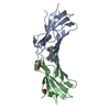


 Mass: 60.095 Da / Num. of mol.: 2 / Source method: obtained synthetically / Formula: C3H8O / Comment: alkaloid*YM
 Mass: 106.120 Da / Num. of mol.: 2 / Source method: obtained synthetically / Formula: C4H10O3
 Mass: 18.015 Da / Num. of mol.: 298 / Source method: isolated from a natural source / Formula: H2O
 Sample preparation
 / Beamline: 23-ID-B / Wavelength: 0.9184
  Processing