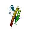
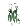
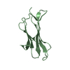
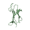
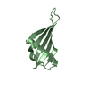
- PDB-2xgl: The X-ray structure of the Escherichia coli colicin M immunity pr... -
+
Open data
ID or keywords:
Loading...
-
Basic information
Entry
Database: PDB / ID: 2xgl
Title
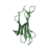
The X-ray structure of the Escherichia coli colicin M immunity protein demonstrates the presence of a disulphide bridge, which is functionally essential
Mass: 18.015 Da / Num. of mol.: 15 / Source method: isolated from a natural source / Formula: H2O
Has protein modification
Y
Sequence details
THE CMI OPEN READING FRAME CONTAINS FOUR PUTATIVE ATG INITIATION CODONS. OLSCHLAGER AND COWORKERS ...THE CMI OPEN READING FRAME CONTAINS FOUR PUTATIVE ATG INITIATION CODONS. OLSCHLAGER AND COWORKERS EARLIER DETERMINED AS MKVIS THE N-TERMINAL AMINO ACID SEQUENCE OF THE PROTEIN EXPRESSED IN VIVO, DESIGNATING THE THIRD ATG AS THE ACTUAL TRANSLATION START SIGNAL (OLSCHLAGER, T., A. TURBA, AND V. BRAUN. 1991. BINDING OF THE IMMUNITY PROTEIN INACTIVATES COLICIN M. MOL MICROBIOL 5,1105-1111.)
-
Experimental details
-
Experiment
Experiment
Method: X-RAY DIFFRACTION / Number of used crystals: 1
-
Sample preparation
Crystal
Density Matthews: 2.33 Å3/Da / Density % sol: 47.12 % / Description: NONE
Crystal grow
pH: 6.5 Details: 100 MM MES BUFFER, PH 6.5, AND 12% PEG 20,000, 90 MM CDSO4
Monochromator: SI CHANNEL CUT MONOCHROMATOR / Protocol: SINGLE WAVELENGTH / Monochromatic (M) / Laue (L): M / Scattering type: x-ray
Radiation wavelength
Wavelength: 1.907 Å / Relative weight: 1
Reflection
Resolution: 2.7→50 Å / Num. obs: 6651 / % possible obs: 99.1 % / Observed criterion σ(I): -3 / Redundancy: 4.9 % / Rmerge(I) obs: 0.1 / Net I/σ(I): 14.3
Reflection shell
Resolution: 2.7→2.8 Å / Redundancy: 3.15 % / Rmerge(I) obs: 0.31 / Mean I/σ(I) obs: 5 / % possible all: 94.2
-
Processing
Software
Name
Version
Classification
REFMAC
5.5.0102
refinement
XDS
datareduction
XSCALE
datascaling
SHARP
phasing
Refinement
Method to determine structure: SAD Starting model: NONE Resolution: 2.7→46.24 Å / Cor.coef. Fo:Fc: 0.898 / Cor.coef. Fo:Fc free: 0.871 / SU B: 15.795 / SU ML: 0.182 / Cross valid method: THROUGHOUT / ESU R: 1.532 / ESU R Free: 0.361 / Stereochemistry target values: MAXIMUM LIKELIHOOD / Details: HYDROGENS HAVE BEEN ADDED IN THE RIDING POSITIONS.
Rfactor
Num. reflection
% reflection
Selection details
Rfree
0.26788
308
4.6 %
RANDOM
Rwork
0.22183
-
-
-
obs
0.22395
6342
99.12 %
-
Solvent computation
Ion probe radii: 0.8 Å / Shrinkage radii: 0.8 Å / VDW probe radii: 1.4 Å / Solvent model: MASK
 Movie
Movie Controller

 Yorodumi
 Open data
 Basic information
 Components
 Keywords
 Function and homology information
 X-RAY DIFFRACTION /
  Authors
 Citation
 Structure visualization
 Downloads & links
 Other downloads
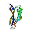

 PDBj
PDBj Assembly
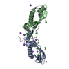

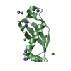

 Mass: 112.411 Da / Num. of mol.: 7 / Source method: obtained synthetically / Formula: Cd
 Mass: 35.453 Da / Num. of mol.: 11 / Source method: obtained synthetically / Formula: Cl
 Mass: 22.990 Da / Num. of mol.: 14 / Source method: obtained synthetically / Formula: Na
 Mass: 18.015 Da / Num. of mol.: 15 / Source method: isolated from a natural source / Formula: H2O
 Sample preparation
 / Beamline: PROXIMA 1 / Wavelength: 1.907
  Processing