 Movie
Movie Controller

[English] 日本語
 Yorodumi
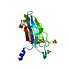
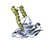
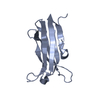
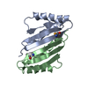
- PDB-1gyw: Gamma-adaptin appendage domain from clathrin adaptor AP1 A753D mutant -
+ Open data

- Basic information
| Entry | Database: PDB / ID: 1gyw | ||||||
|---|---|---|---|---|---|---|---|
| Title | Gamma-adaptin appendage domain from clathrin adaptor AP1 A753D mutant | ||||||
 Components Components | ADAPTER-RELATED PROTEIN COMPLEX 1 GAMMA 1 SUBUNIT | ||||||
 Keywords Keywords | ENDOCYTOSIS / ADAPTOR / COATED PITS | ||||||
| Function / homology |  Function and homology information Function and homology informationbasolateral protein secretion / synaptic vesicle budding from endosome / Lysosome Vesicle Biogenesis / positive regulation of natural killer cell degranulation / endosome to melanosome transport / AP-1 adaptor complex / platelet dense granule organization / melanosome assembly / Golgi to lysosome transport / Golgi to vacuole transport ...basolateral protein secretion / synaptic vesicle budding from endosome / Lysosome Vesicle Biogenesis / positive regulation of natural killer cell degranulation / endosome to melanosome transport / AP-1 adaptor complex / platelet dense granule organization / melanosome assembly / Golgi to lysosome transport / Golgi to vacuole transport / Golgi Associated Vesicle Biogenesis / clathrin-cargo adaptor activity / MHC class II antigen presentation / GTP-dependent protein binding / positive regulation of natural killer cell mediated cytotoxicity / clathrin-coated vesicle / kinesin binding / synaptic vesicle endocytosis / collagen binding / vesicle-mediated transport / clathrin-coated pit / trans-Golgi network membrane / trans-Golgi network / intracellular protein transport / recycling endosome / small GTPase binding / presynapse / early endosome / lysosomal membrane / perinuclear region of cytoplasm / Golgi apparatus / cytosol Similarity search - Function | ||||||
| Biological species |  | ||||||
| Method |  X-RAY DIFFRACTION / MOLECULAR REPLACEMENT / Resolution: 2.4 Å X-RAY DIFFRACTION / MOLECULAR REPLACEMENT / Resolution: 2.4 Å | ||||||
 Authors Authors | Evans, P.R. / Miele, A.E. / Owen, D.J. / McMahon, H.M. / Kent, H.M. | ||||||
 Citation Citation | Journal: Structure / Year: 2002 Title: Gamma-adaptin appendage domain: structure and binding site for Eps15 and gamma-synergin. Authors: Kent, H.M. / McMahon, H.T. / Evans, P.R. / Benmerah, A. / Owen, D.J. | ||||||
| History |
|
- Structure visualization
| Structure viewer | Molecule: MolmilJmol/JSmol |
|---|
- Downloads & links
-Download
| PDBx/mmCIF format | 1gyw.cif.gz | 64.2 KB | Display | PDBx/mmCIF format |
|---|---|---|---|---|
| PDB format | pdb1gyw.ent.gz | 48 KB | Display | PDB format |
| PDBx/mmJSON format | 1gyw.json.gz | Tree view | PDBx/mmJSON format | |
| Others |  Other downloads Other downloads |
-Validation report
| Arichive directory | https://data.pdbj.org/pub/pdb/validation_reports/gy/1gywftp://data.pdbj.org/pub/pdb/validation_reports/gy/1gyw | HTTPS FTP |
|---|
-Related structure data
| Related structure data |  1gyuSC  1gyvC S: Starting model for refinement C: citing same article ( |
|---|---|
| Similar structure data |
-Links
 PDBj
PDBj


- Assembly
| Deposited unit | 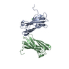
| ||||||||
|---|---|---|---|---|---|---|---|---|---|
| 1 | 
| ||||||||
| 2 | 
| ||||||||
| Unit cell |
| ||||||||
| Noncrystallographic symmetry (NCS) | NCS oper: (Code: given Matrix: (0.134391, 0.511664, 0.84861), Vector: |
-Components
| #1: Protein | Mass: 14255.128 Da / Num. of mol.: 2 / Fragment: APPENDAGE DOMAIN, RESIDUES 694-821 / Mutation: YES Source method: isolated from a genetically manipulated source Source: (gene. exp.)  #2: Chemical | ChemComp-CL / |   Mass: 35.453 Da / Num. of mol.: 1 / Source method: obtained synthetically / Formula: Cl Mass: 35.453 Da / Num. of mol.: 1 / Source method: obtained synthetically / Formula: Cl#3: Water | ChemComp-HOH / |  Mass: 18.015 Da / Num. of mol.: 178 / Source method: isolated from a natural source / Formula: H2O Mass: 18.015 Da / Num. of mol.: 178 / Source method: isolated from a natural source / Formula: H2O |
|---|
-Experimental details
-Experiment
| Experiment | Method: X-RAY DIFFRACTION / Number of used crystals: 1 |
|---|
- Sample preparation
| Crystal | Density Matthews: 2.52 Å3/Da / Density % sol: 55.9 % | ||||||||||||||||||||||||||||||||||||||||||||||||||||||||
|---|---|---|---|---|---|---|---|---|---|---|---|---|---|---|---|---|---|---|---|---|---|---|---|---|---|---|---|---|---|---|---|---|---|---|---|---|---|---|---|---|---|---|---|---|---|---|---|---|---|---|---|---|---|---|---|---|---|
| Crystal grow | Method: vapor diffusion / pH: 8 Details: 24% PEG4000, 100 MM TRIS PH 8.0, 200MM MGCL2, VAPOUR DIFFUSION | ||||||||||||||||||||||||||||||||||||||||||||||||||||||||
| Crystal grow | *PLUS Method: vapor diffusion, hanging drop | ||||||||||||||||||||||||||||||||||||||||||||||||||||||||
| Components of the solutions | *PLUS
|
-Data collection
| Diffraction | Mean temperature: 100 K |
|---|---|
| Diffraction source | Source: ROTATING ANODE / Type: RIGAKU RU200 / Wavelength: 1.54182 |
| Detector | Type: MARRESEARCH / Detector: IMAGE PLATE / Date: Jul 15, 2001 / Details: MIRRORS |
| Radiation | Protocol: SINGLE WAVELENGTH / Monochromatic (M) / Laue (L): M / Scattering type: x-ray |
| Radiation wavelength | Wavelength: 1.54182 Å / Relative weight: 1 |
| Reflection | Resolution: 2.4→23.06 Å / Num. obs: 11979 / % possible obs: 99.3 % / Observed criterion σ(I): 6 / Redundancy: 13.62 % / Rmerge(I) obs: 0.086 / Net I/σ(I): 8.1922 |
| Reflection shell | Resolution: 2.4→2.53 Å / Redundancy: 13.41 % / Rmerge(I) obs: 0.33 / Mean I/σ(I) obs: 2.18 / % possible all: 96.3 |
- Processing
| Software |
| ||||||||||||||||||||
|---|---|---|---|---|---|---|---|---|---|---|---|---|---|---|---|---|---|---|---|---|---|
| Refinement | Method to determine structure: MOLECULAR REPLACEMENT Starting model: WILD-TYPE 1GYU Resolution: 2.4→57.74 Å / SU B: 7.025 / SU ML: 0.166 / Cross valid method: THROUGHOUT / ESU R Free: 0.288 / Details: NONE
| ||||||||||||||||||||
| Displacement parameters | Biso mean: 27.773 Å2
| ||||||||||||||||||||
| Refinement step | Cycle: LAST / Resolution: 2.4→57.74 Å
| ||||||||||||||||||||
| LS refinement shell | *PLUS Highest resolution: 2.404 Å / Lowest resolution: 2.467 Å / Rfactor Rfree: 0.266 / Rfactor Rwork: 0.235 / Total num. of bins used: 20 |
