 Movie
Movie Controller

[English] 日本語
 Yorodumi
- PDB-1rsy: STRUCTURE OF THE FIRST C2-DOMAIN OF SYNAPTOTAGMIN I: A NOVEL CA2+... -
+ Open data

- Basic information
| Entry | Database: PDB / ID: 1rsy | ||||||
|---|---|---|---|---|---|---|---|
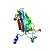
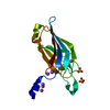
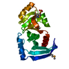
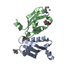
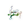
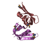
| Title | STRUCTURE OF THE FIRST C2-DOMAIN OF SYNAPTOTAGMIN I: A NOVEL CA2+(SLASH)PHOSPHOLIPID BINDING FOLD | ||||||
 Components Components | SYNAPTOTAGMIN I | ||||||
 Keywords Keywords | CALCIUM/PHOSPHOLIPID BINDING PROTEIN / CALCIUM-PHOSPHOLIPID BINDING PROTEIN complex | ||||||
| Function / homology |  Function and homology information Function and homology informationsynchronous neurotransmitter secretion / regulation of vesicle fusion / spontaneous neurotransmitter secretion / fast, calcium ion-dependent exocytosis of neurotransmitter / syntaxin-3 binding / regulation of regulated secretory pathway / regulation of calcium-dependent activation of synaptic vesicle fusion / calcium-dependent activation of synaptic vesicle fusion / chromaffin granule membrane / : ...synchronous neurotransmitter secretion / regulation of vesicle fusion / spontaneous neurotransmitter secretion / fast, calcium ion-dependent exocytosis of neurotransmitter / syntaxin-3 binding / regulation of regulated secretory pathway / regulation of calcium-dependent activation of synaptic vesicle fusion / calcium-dependent activation of synaptic vesicle fusion / chromaffin granule membrane / : / positive regulation of calcium ion-dependent exocytosis of neurotransmitter / calcium activated phospholipid scrambling / calcium ion-regulated exocytosis of neurotransmitter / Glutamate Neurotransmitter Release Cycle / Norepinephrine Neurotransmitter Release Cycle / Acetylcholine Neurotransmitter Release Cycle / Serotonin Neurotransmitter Release Cycle / GABA synthesis, release, reuptake and degradation / regulation of calcium ion-dependent exocytosis / Dopamine Neurotransmitter Release Cycle / calcium ion sensor activity / synaptic vesicle recycling / calcium-ion regulated exocytosis / exocytic vesicle / positive regulation of calcium ion-dependent exocytosis / protein heterooligomerization / vesicle organization / : / positive regulation of vesicle fusion / positive regulation of dendrite extension / Cargo recognition for clathrin-mediated endocytosis / Clathrin-mediated endocytosis / vesicle fusion / calcium-dependent phospholipid binding / positive regulation of dopamine secretion / negative regulation of neurotransmitter secretion / dense core granule / membraneless organelle assembly / xenobiotic transmembrane transport / neurotransmitter secretion / syntaxin binding / syntaxin-1 binding / presynaptic active zone / inhibitory postsynaptic potential / low-density lipoprotein particle receptor binding / phosphatidylserine binding / clathrin binding / regulation of synaptic vesicle exocytosis / neuron projection terminus / regulation of dopamine secretion / protein insertion into membrane / regulation of endocytosis / synaptic vesicle exocytosis / detection of calcium ion / postsynaptic cytosol / positive regulation of synaptic transmission / regulation of synaptic transmission, glutamatergic / presynaptic cytosol / vesicle-mediated transport / phosphatidylinositol-4,5-bisphosphate binding / excitatory synapse / secretory granule / cellular response to calcium ion / hippocampal mossy fiber to CA3 synapse / SNARE binding / molecular condensate scaffold activity / phospholipid binding / response to calcium ion / calcium-dependent protein binding / terminal bouton / synaptic vesicle / synaptic vesicle membrane / presynaptic membrane / cell differentiation / calmodulin binding / postsynaptic membrane / neuron projection / postsynaptic density / protein heterodimerization activity / axon / calcium ion binding / lipid binding / glutamatergic synapse / Golgi apparatus / identical protein binding / plasma membrane / cytoplasm Similarity search - Function | ||||||
| Biological species |  | ||||||
| Method |  X-RAY DIFFRACTION / Resolution: 1.9 Å X-RAY DIFFRACTION / Resolution: 1.9 Å | ||||||
 Authors Authors | Sutton, R.B. / Sprang, S.R. | ||||||
 Citation Citation | Journal: Cell(Cambridge,Mass.) / Year: 1995 Title: Structure of the first C2 domain of synaptotagmin I: a novel Ca2+/phospholipid-binding fold. Authors: Sutton, R.B. / Davletov, B.A. / Berghuis, A.M. / Sudhof, T.C. / Sprang, S.R. #1: Journal: J.Biol.Chem. / Year: 1993Title: A Single C2 Domain from Synaptotagmin I is Sufficient for High Affinity Ca2+(Slash)Phospholipid Binding Authors: Davletov, B.A. / Sudhof, T.C. | ||||||
| History |
|
- Structure visualization
| Structure viewer | Molecule: MolmilJmol/JSmol |
|---|
- Downloads & links
-Download
| PDBx/mmCIF format | 1rsy.cif.gz | 40.9 KB | Display | PDBx/mmCIF format |
|---|---|---|---|---|
| PDB format | pdb1rsy.ent.gz | 27.5 KB | Display | PDB format |
| PDBx/mmJSON format | 1rsy.json.gz | Tree view | PDBx/mmJSON format | |
| Others |  Other downloads Other downloads |
-Validation report
| Arichive directory | https://data.pdbj.org/pub/pdb/validation_reports/rs/1rsyftp://data.pdbj.org/pub/pdb/validation_reports/rs/1rsy | HTTPS FTP |
|---|
-Related structure data
| Similar structure data |
|---|


-Links
 PDBj
PDBj


- Assembly
| Deposited unit | 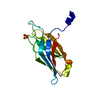
| ||||||||
|---|---|---|---|---|---|---|---|---|---|
| 1 |
| ||||||||
| Unit cell |
| ||||||||
| Atom site foot note | 1: CIS PROLINE - PRO 187 |
-Components
| #1: Protein | Mass: 17098.350 Da / Num. of mol.: 1 Source method: isolated from a genetically manipulated source Source: (gene. exp.)  |
|---|---|
| #2: Chemical | ChemComp-SO4 /   Mass: 96.063 Da / Num. of mol.: 1 / Source method: obtained synthetically / Formula: SO4 Mass: 96.063 Da / Num. of mol.: 1 / Source method: obtained synthetically / Formula: SO4 |
| #3: Water | ChemComp-HOH /  Mass: 18.015 Da / Num. of mol.: 80 / Source method: isolated from a natural source / Formula: H2O Mass: 18.015 Da / Num. of mol.: 80 / Source method: isolated from a natural source / Formula: H2O |
| Nonpolymer details | HET GROUP TRIVIAL NAME: SULFATE EMPIRICAL FORMULA: SO4 NUMBER OF ATOMS IN GROUP: 5 ADDITIONAL ...HET GROUP TRIVIAL NAME: SULFATE EMPIRICAL FORMULA: SO4 NUMBER OF ATOMS IN GROUP: 5 ADDITIONAL |
-Experimental details
-Experiment
| Experiment | Method: X-RAY DIFFRACTION |
|---|
- Sample preparation
| Crystal | Density Matthews: 2.13 Å3/Da / Density % sol: 42.24 % | ||||||||||||||||||||||||
|---|---|---|---|---|---|---|---|---|---|---|---|---|---|---|---|---|---|---|---|---|---|---|---|---|---|
| Crystal | *PLUS Density % sol: 40 % | ||||||||||||||||||||||||
| Crystal grow | *PLUS Temperature: 21 ℃ / pH: 7.2 / Method: vapor diffusion, hanging drop | ||||||||||||||||||||||||
| Components of the solutions | *PLUS
|
-Data collection
| Diffraction source | Wavelength: 1.5418 |
|---|---|
| Detector | Type: RIGAKU RAXIS IIC / Detector: IMAGE PLATE |
| Radiation | Monochromatic (M) / Laue (L): M / Scattering type: x-ray |
| Radiation wavelength | Wavelength: 1.5418 Å / Relative weight: 1 |
| Reflection | Num. obs: 11214 / % possible obs: 95 % / Observed criterion σ(I): 2 / Redundancy: 2.8 % |
- Processing
| Software |
| ||||||||||||||||||||||||||||||||||||||||||||||||||||||||||||
|---|---|---|---|---|---|---|---|---|---|---|---|---|---|---|---|---|---|---|---|---|---|---|---|---|---|---|---|---|---|---|---|---|---|---|---|---|---|---|---|---|---|---|---|---|---|---|---|---|---|---|---|---|---|---|---|---|---|---|---|---|---|
| Refinement | Resolution: 1.9→6 Å / σ(F): 2
| ||||||||||||||||||||||||||||||||||||||||||||||||||||||||||||
| Displacement parameters | Biso mean: 24 Å2 | ||||||||||||||||||||||||||||||||||||||||||||||||||||||||||||
| Refinement step | Cycle: LAST / Resolution: 1.9→6 Å
| ||||||||||||||||||||||||||||||||||||||||||||||||||||||||||||
| Refine LS restraints |
| ||||||||||||||||||||||||||||||||||||||||||||||||||||||||||||
| Refinement | *PLUS | ||||||||||||||||||||||||||||||||||||||||||||||||||||||||||||
| Solvent computation | *PLUS | ||||||||||||||||||||||||||||||||||||||||||||||||||||||||||||
| Displacement parameters | *PLUS | ||||||||||||||||||||||||||||||||||||||||||||||||||||||||||||
| Refine LS restraints | *PLUS
|
