 Movie
Movie Controller

[English] 日本語
 Yorodumi
- PDB-3zry: Rotor architecture in the F(1)-c(10)-ring complex of the yeast F-... -
+ Open data

- Basic information
| Entry | Database: PDB / ID: 3zry | |||||||||
|---|---|---|---|---|---|---|---|---|---|---|
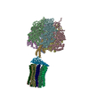
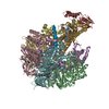
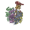
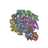
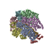
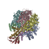
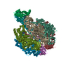
| Title | Rotor architecture in the F(1)-c(10)-ring complex of the yeast F-ATP synthase | |||||||||
 Components Components |
| |||||||||
 Keywords Keywords |  HYDROLASE / ATP-BINDING / F(1)-F(O)ATP SYNTHASE / MITOCHONDRIA / MOLECULAR MOTOR / CENTRAL STALK / MEMBRANE PROTEIN / C-RING HYDROLASE / ATP-BINDING / F(1)-F(O)ATP SYNTHASE / MITOCHONDRIA / MOLECULAR MOTOR / CENTRAL STALK / MEMBRANE PROTEIN / C-RING | |||||||||
| Function / homology |  Function and homology information Function and homology informationmitochondrial proton-transporting ATP synthase, central stalk / mitochondrial proton-transporting ATP synthase, catalytic core / mitochondrial proton-transporting ATP synthase complex, coupling factor F(o) / mitochondrial proton-transporting ATP synthase complex / mitochondrial proton-transporting ATP synthase complex, catalytic sector F(1) / mitochondrial nucleoid / proton motive force-driven mitochondrial ATP synthesis / proton motive force-driven ATP synthesis / proton transmembrane transporter activity / proton-transporting ATP synthase complex, catalytic core F(1) ...mitochondrial proton-transporting ATP synthase, central stalk / mitochondrial proton-transporting ATP synthase, catalytic core / mitochondrial proton-transporting ATP synthase complex, coupling factor F(o) / mitochondrial proton-transporting ATP synthase complex / mitochondrial proton-transporting ATP synthase complex, catalytic sector F(1) / mitochondrial nucleoid / proton motive force-driven mitochondrial ATP synthesis / proton motive force-driven ATP synthesis / proton transmembrane transporter activity / proton-transporting ATP synthase complex, catalytic core F(1) / H+-transporting two-sector ATPase / proton-transporting ATPase activity, rotational mechanism / proton-transporting ATP synthase activity, rotational mechanism / ADP binding / mitochondrial intermembrane space / mitochondrial inner membrane / lipid binding / ATP hydrolysis activity / mitochondrion / ATP binding / identical protein binding / cytosolSimilarity search - Function | |||||||||
| Biological species |  SACCHAROMYCES CEREVISIAE (brewer's yeast) SACCHAROMYCES CEREVISIAE (brewer's yeast) | |||||||||
| Method | X-RAY DIFFRACTION / SYNCHROTRON / MOLECULAR REPLACEMENT / Resolution: 6.5 Å | |||||||||
 Authors Authors | Giraud, M.-F. / Dautant, A. | |||||||||
 Citation Citation | Journal: J.Struct.Biol. / Year: 2012 Title: Rotor Architecture in the Yeast and Bovine F(1)-C-Ring Complexes of F-ATP Synthase. Authors: Giraud, M.-F. / Paumard, P. / Sanchez, C. / Brethes, D. / Velours, J. / Dautant, A. #1: Journal: J.Biol.Chem. / Year: 2010Title: Crystal Structure of the Mgadp-Inhibited State of the Yeast F1C10-ATP Synthase. Authors: Dautant, A. / Velours, J. / Giraud, M. #2: Journal: J.Bioenerg.Biomembr. / Year: 2009 Title: Hydrogenated and Fluorinated Surfactants Derived from Tris(Hydroxymethyl)-Acrylamidomethane Allow the Purification of a Highly Active Yeast F1-F0 ATP-Synthase with an Enhanced Stability. Authors: Talbot, J.-C. / Dautant, A. / Polidori, A. / Pucci, B. / Cohen-Bouhacina, T. / Maali, A. / Salin, B. / Brethes, D. / Velours, J. / Giraud, M.-F. #3: Journal: Science / Year: 1999Title: Molecular Architecture of the Rotary Motor in ATP Synthase Authors: Stock, D. / Leslie, A.G.W. / Walker, J.E. #4: Journal: Embo J. / Year: 2006Title: Novel Features of the Rotary Catalytic Mechanism Revealed in the Structure of Yeast F1 ATPase Authors: Kabaleeswaran, V. / Puri, N. / Walker, J.E. / Leslie, A.G. / Mueller, D.M. | |||||||||
| History |
| |||||||||
| Remark 700 | SHEET DETERMINATION METHOD: DSSP THE SHEETS PRESENTED AS "AA" IN EACH CHAIN ON SHEET RECORDS BELOW ... SHEET DETERMINATION METHOD: DSSP THE SHEETS PRESENTED AS "AA" IN EACH CHAIN ON SHEET RECORDS BELOW IS ACTUALLY AN 0-STRANDED BARREL THIS IS REPRESENTED BY A 1-STRANDED SHEET IN WHICH THE FIRST AND LAST STRANDS ARE IDENTICAL. THE SHEETS PRESENTED AS "BA" IN EACH CHAIN ON SHEET RECORDS BELOW IS ACTUALLY AN 6-STRANDED BARREL THIS IS REPRESENTED BY A 7-STRANDED SHEET IN WHICH THE FIRST AND LAST STRANDS ARE IDENTICAL. THE SHEETS PRESENTED AS "EA" IN EACH CHAIN ON SHEET RECORDS BELOW IS ACTUALLY AN 6-STRANDED BARREL THIS IS REPRESENTED BY A 7-STRANDED SHEET IN WHICH THE FIRST AND LAST STRANDS ARE IDENTICAL. THE SHEETS PRESENTED AS "FA" IN EACH CHAIN ON SHEET RECORDS BELOW IS ACTUALLY AN 6-STRANDED BARREL THIS IS REPRESENTED BY A 7-STRANDED SHEET IN WHICH THE FIRST AND LAST STRANDS ARE IDENTICAL. |
- Structure visualization
| Structure viewer | Molecule: MolmilJmol/JSmol |
|---|
- Downloads & links
-Download
| PDBx/mmCIF format | 3zry.cif.gz | 744 KB | Display | PDBx/mmCIF format |
|---|---|---|---|---|
| PDB format | pdb3zry.ent.gz | 606.5 KB | Display | PDB format |
| PDBx/mmJSON format | 3zry.json.gz | Tree view | PDBx/mmJSON format | |
| Others |  Other downloads Other downloads |
-Validation report
| Arichive directory | https://data.pdbj.org/pub/pdb/validation_reports/zr/3zryftp://data.pdbj.org/pub/pdb/validation_reports/zr/3zry | HTTPS FTP |
|---|
-Related structure data
| Related structure data |  2xokS S: Starting model for refinement |
|---|---|
| Similar structure data |


-Links
 PDBj
PDBj


- Assembly
| Deposited unit | 
| ||||||||||||||||||||||||||||||||||||||||||||||
|---|---|---|---|---|---|---|---|---|---|---|---|---|---|---|---|---|---|---|---|---|---|---|---|---|---|---|---|---|---|---|---|---|---|---|---|---|---|---|---|---|---|---|---|---|---|---|---|
| 1 | 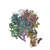
| ||||||||||||||||||||||||||||||||||||||||||||||
| 2 | 
| ||||||||||||||||||||||||||||||||||||||||||||||
| Unit cell |
| ||||||||||||||||||||||||||||||||||||||||||||||
| Noncrystallographic symmetry (NCS) | NCS domain:
NCS domain segments:
|