 Movie
Movie Controller

[English] 日本語
 Yorodumi
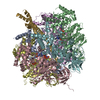
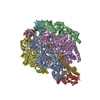
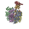
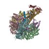
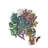
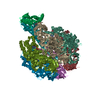
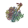
- PDB-4yxw: Bovine heart mitochondrial F1-ATPase inhibited by AMP-PNP and ADP... -
+ Open data

- Basic information
| Entry | Database: PDB / ID: 4yxw | ||||||
|---|---|---|---|---|---|---|---|
| Title | Bovine heart mitochondrial F1-ATPase inhibited by AMP-PNP and ADP in the presence of thiophosphate. | ||||||
 Components Components | (ATP synthase subunit ...) x 5 | ||||||
 Keywords Keywords | HYDROLASE / complex / mitochondrial | ||||||
| Function / homology |  Function and homology information Function and homology informationFormation of ATP by chemiosmotic coupling / Cristae formation / mitochondrial proton-transporting ATP synthase complex assembly / mitochondrial envelope / Mitochondrial protein degradation / proton transmembrane transporter activity / proton motive force-driven ATP synthesis / proton motive force-driven mitochondrial ATP synthesis / H+-transporting two-sector ATPase / proton-transporting ATP synthase complex ...Formation of ATP by chemiosmotic coupling / Cristae formation / mitochondrial proton-transporting ATP synthase complex assembly / mitochondrial envelope / Mitochondrial protein degradation / proton transmembrane transporter activity / proton motive force-driven ATP synthesis / proton motive force-driven mitochondrial ATP synthesis / H+-transporting two-sector ATPase / proton-transporting ATP synthase complex / proton-transporting ATP synthase activity, rotational mechanism / proton transmembrane transport / aerobic respiration / ADP binding / mitochondrial inner membrane / structural molecule activity / ATP hydrolysis activity / mitochondrion / ATP binding / metal ion binding / plasma membrane Similarity search - Function | ||||||
| Biological species |  | ||||||
| Method |  X-RAY DIFFRACTION / SYNCHROTRON / MOLECULAR REPLACEMENT / molecular replacement / Resolution: 3.1 Å X-RAY DIFFRACTION / SYNCHROTRON / MOLECULAR REPLACEMENT / molecular replacement / Resolution: 3.1 Å | ||||||
 Authors Authors | Bason, J.V. / Montgomery, M.G. / Leslie, A.G.W. / Walker, J.E. | ||||||
 Citation Citation | Journal: Proc.Natl.Acad.Sci.USA / Year: 2015 Title: How release of phosphate from mammalian F1-ATPase generates a rotary substep. Authors: Bason, J.V. / Montgomery, M.G. / Leslie, A.G. / Walker, J.E. | ||||||
| History |
|
- Structure visualization
| Structure viewer | Molecule: MolmilJmol/JSmol |
|---|
- Downloads & links
-Download
| PDBx/mmCIF format | 4yxw.cif.gz | 617.7 KB | Display | PDBx/mmCIF format |
|---|---|---|---|---|
| PDB format | pdb4yxw.ent.gz | 501.2 KB | Display | PDB format |
| PDBx/mmJSON format | 4yxw.json.gz | Tree view | PDBx/mmJSON format | |
| Others |  Other downloads Other downloads |
-Validation report
| Arichive directory | https://data.pdbj.org/pub/pdb/validation_reports/yx/4yxwftp://data.pdbj.org/pub/pdb/validation_reports/yx/4yxw | HTTPS FTP |
|---|
-Related structure data
| Related structure data |  4z1mC 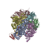 2ck3S S: Starting model for refinement C: citing same article ( |
|---|---|
| Similar structure data |


-Links
 PDBj
PDBj


- Assembly
| Deposited unit | 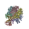
| ||||||||
|---|---|---|---|---|---|---|---|---|---|
| 1 |
| ||||||||
| Unit cell |
|
-Components
-ATP synthase subunit ... , 5 types, 9 molecules ABCDEFGHI
| #1: Protein | Mass: 55302.191 Da / Num. of mol.: 3 / Source method: isolated from a natural source / Source: (natural) #2: Protein | Mass: 51757.836 Da / Num. of mol.: 3 / Source method: isolated from a natural source / Source: (natural) References: UniProt: P00829, H+-transporting two-sector ATPase #3: Protein | | Mass: 30300.760 Da / Num. of mol.: 1 / Source method: isolated from a natural source / Source: (natural) #4: Protein | | Mass: 15074.813 Da / Num. of mol.: 1 / Source method: isolated from a natural source / Source: (natural) #5: Protein/peptide | | Mass: 5662.693 Da / Num. of mol.: 1 / Source method: isolated from a natural source / Source: (natural) |
|---|
-Non-polymers , 6 types, 50 molecules 


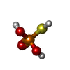


| #6: Chemical | ChemComp-ANP /  Mass: 506.196 Da / Num. of mol.: 5 / Source method: obtained synthetically / Formula: C10H17N6O12P3 / Comment: AMP-PNP, energy-carrying molecule analogue*YM Mass: 506.196 Da / Num. of mol.: 5 / Source method: obtained synthetically / Formula: C10H17N6O12P3 / Comment: AMP-PNP, energy-carrying molecule analogue*YM#7: Chemical | ChemComp-MG /  Mass: 24.305 Da / Num. of mol.: 5 / Source method: obtained synthetically / Formula: Mg Mass: 24.305 Da / Num. of mol.: 5 / Source method: obtained synthetically / Formula: Mg#8: Chemical | ChemComp-CL / |  Mass: 35.453 Da / Num. of mol.: 1 / Source method: obtained synthetically / Formula: Cl Mass: 35.453 Da / Num. of mol.: 1 / Source method: obtained synthetically / Formula: Cl#9: Chemical | ChemComp-TS6 / |  Mass: 114.061 Da / Num. of mol.: 1 / Source method: obtained synthetically / Formula: H3O3PS Mass: 114.061 Da / Num. of mol.: 1 / Source method: obtained synthetically / Formula: H3O3PS#10: Chemical | ChemComp-NA / |  Mass: 22.990 Da / Num. of mol.: 1 / Source method: obtained synthetically / Formula: Na Mass: 22.990 Da / Num. of mol.: 1 / Source method: obtained synthetically / Formula: Na#11: Water | ChemComp-HOH / | Mass: 18.015 Da / Num. of mol.: 37 / Source method: isolated from a natural source / Formula: H2O |
|---|
-Details
| Compound details | SER 481 GLY in chains A, B and C was identified as a GLY from the protein sequence. In the cDNA ...SER 481 GLY in chains A, B and C was identified as a GLY from the protein sequence. In the cDNA sequence, the codon for this residue was AGC SER in three clones while in two others it was GGC GLY. The difference was thought to be due to a mutation occurring during either propagation of the clones in the library or subcloning into M13 vectors. The electron density suggests a Gly in this position. |
|---|
-Experimental details
-Experiment
| Experiment | Method: X-RAY DIFFRACTION / Number of used crystals: 1 |
|---|
- Sample preparation
| Crystal | Density Matthews: 2.27 Å3/Da / Density % sol: 45.92 % |
|---|---|
| Crystal grow | Temperature: 295 K / Method: microdialysis / pH: 8.2 Details: PEG 6000, sodium chloride, magnesium chloride, Tris-HCl, AMP-PNP, ADP, sodium monothiophosphate |
-Data collection
| Diffraction | Mean temperature: 100 K | ||||||||||||||||||||||||
|---|---|---|---|---|---|---|---|---|---|---|---|---|---|---|---|---|---|---|---|---|---|---|---|---|---|
| Diffraction source | Source: SYNCHROTRON / Site: Diamond  / Beamline: I02 / Wavelength: 0.979 Å / Beamline: I02 / Wavelength: 0.979 Å | ||||||||||||||||||||||||
| Detector | Type: DECTRIS PILATUS 6M / Detector: PIXEL / Date: Aug 6, 2014 | ||||||||||||||||||||||||
| Radiation | Protocol: SINGLE WAVELENGTH / Monochromatic (M) / Laue (L): M / Scattering type: x-ray | ||||||||||||||||||||||||
| Radiation wavelength | Wavelength: 0.979 Å / Relative weight: 1 | ||||||||||||||||||||||||
| Reflection | Resolution: 3.1→76.52 Å / Num. obs: 59186 / % possible obs: 95.4 % / Redundancy: 2.6 % / Biso Wilson estimate: 53.48 Å2 / CC1/2: 0.884 / Rmerge(I) obs: 0.171 / Rpim(I) all: 0.123 / Net I/σ(I): 6.4 / Num. measured all: 154361 / Scaling rejects: 36 | ||||||||||||||||||||||||
| Reflection shell | Diffraction-ID: 1 / Redundancy: 2.7 % / Rejects: _
|
-Phasing
| Phasing | Method: molecular replacement |
|---|
- Processing
| Software |
| |||||||||||||||||||||||||||||||||||||||||||||||||||||||||||||||||||||||||||
|---|---|---|---|---|---|---|---|---|---|---|---|---|---|---|---|---|---|---|---|---|---|---|---|---|---|---|---|---|---|---|---|---|---|---|---|---|---|---|---|---|---|---|---|---|---|---|---|---|---|---|---|---|---|---|---|---|---|---|---|---|---|---|---|---|---|---|---|---|---|---|---|---|---|---|---|---|
| Refinement | Method to determine structure: MOLECULAR REPLACEMENT Starting model: 2CK3 Resolution: 3.1→76.5 Å / Cor.coef. Fo:Fc: 0.913 / Cor.coef. Fo:Fc free: 0.883 / WRfactor Rfree: 0.2773 / WRfactor Rwork: 0.2283 / FOM work R set: 0.7774 / SU B: 28.758 / SU ML: 0.488 / SU Rfree: 0.5867 / Cross valid method: THROUGHOUT / σ(F): 0 / ESU R Free: 0.587 / Stereochemistry target values: MAXIMUM LIKELIHOOD Details: HYDROGENS HAVE BEEN ADDED IN THE RIDING POSITIONS U VALUES : REFINED INDIVIDUALLY
| |||||||||||||||||||||||||||||||||||||||||||||||||||||||||||||||||||||||||||
| Solvent computation | Ion probe radii: 0.8 Å / Shrinkage radii: 0.8 Å / VDW probe radii: 1.2 Å / Solvent model: MASK | |||||||||||||||||||||||||||||||||||||||||||||||||||||||||||||||||||||||||||
| Displacement parameters | Biso max: 195.85 Å2 / Biso mean: 75.741 Å2 / Biso min: 24.2 Å2
| |||||||||||||||||||||||||||||||||||||||||||||||||||||||||||||||||||||||||||
| Refinement step | Cycle: final / Resolution: 3.1→76.5 Å
| |||||||||||||||||||||||||||||||||||||||||||||||||||||||||||||||||||||||||||
| Refine LS restraints |
| |||||||||||||||||||||||||||||||||||||||||||||||||||||||||||||||||||||||||||
| LS refinement shell | Resolution: 3.1→3.18 Å / Total num. of bins used: 20
|
