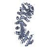
 Movie
Movie Controller


+ Open data

- Basic information
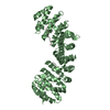
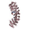
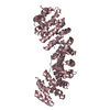
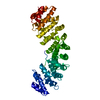
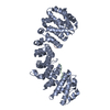
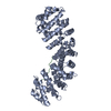
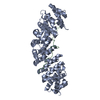
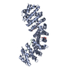
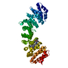
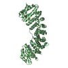
| Entry | Database: PDB / ID: 3zin | ||||||
|---|---|---|---|---|---|---|---|
| Title | Gu_alpha_helicase | ||||||
 Components Components |
| ||||||
 Keywords Keywords | TRANSPORT PROTEIN/HYDROLASE / TRANSPORT PROTEIN-HYDROLASE COMPLEX | ||||||
| Function / homology |  Function and homology information Function and homology informationB-WICH complex positively regulates rRNA expression / Major pathway of rRNA processing in the nucleolus and cytosol / 7SK snRNA binding / R-loop processing / positive regulation of myeloid dendritic cell cytokine production / Sensing of DNA Double Strand Breaks / regulation of transcription by glucose / entry of viral genome into host nucleus through nuclear pore complex via importin / positive regulation of viral life cycle / B-WICH complex ...B-WICH complex positively regulates rRNA expression / Major pathway of rRNA processing in the nucleolus and cytosol / 7SK snRNA binding / R-loop processing / positive regulation of myeloid dendritic cell cytokine production / Sensing of DNA Double Strand Breaks / regulation of transcription by glucose / entry of viral genome into host nucleus through nuclear pore complex via importin / positive regulation of viral life cycle / B-WICH complex / NLS-dependent protein nuclear import complex / postsynapse to nucleus signaling pathway / NLS-bearing protein import into nucleus / nuclear import signal receptor activity / miRNA binding / nuclear localization sequence binding / positive regulation of transcription by RNA polymerase III / non-canonical NF-kappaB signal transduction / snoRNA binding / response to exogenous dsRNA / positive regulation of transcription by RNA polymerase I / positive regulation of type I interferon production / response to virus / histone deacetylase binding / protein import into nucleus / cytoplasmic stress granule / rRNA processing / double-stranded RNA binding / host cell / nuclear membrane / defense response to virus / DNA-binding transcription factor binding / transcription by RNA polymerase II / RNA helicase activity / positive regulation of canonical NF-kappaB signal transduction / rRNA binding / postsynaptic density / RNA helicase / chromatin remodeling / innate immune response / positive regulation of DNA-templated transcription / nucleolus / glutamatergic synapse / positive regulation of transcription by RNA polymerase II / ATP hydrolysis activity / mitochondrion / RNA binding / nucleoplasm / ATP binding / identical protein binding / nucleus / cytoplasm / cytosol Similarity search - Function | ||||||
| Biological species |  | ||||||
| Method |  X-RAY DIFFRACTION / SYNCHROTRON / MOLECULAR REPLACEMENT / Resolution: 2 Å X-RAY DIFFRACTION / SYNCHROTRON / MOLECULAR REPLACEMENT / Resolution: 2 Å | ||||||
 Authors Authors | Chang, C.-W. / Counago, R.M. / Williams, S.J. / Kobe, B. | ||||||
 Citation Citation | Journal: Traffic / Year: 2013 Title: Distinctive Conformation of Minor Site-Specific Nuclear Localization Signals Bound to Importin-Alpha Authors: Chang, C.-W. / Counago, R.M. / Williams, S.J. / Boden, M. / Kobe, B. | ||||||
| History |
|
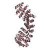
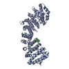
- Structure visualization
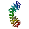
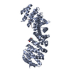
| Structure viewer | Molecule: MolmilJmol/JSmol |
|---|
- Downloads & links
-Download
| PDBx/mmCIF format | 3zin.cif.gz | 182.3 KB | Display | PDBx/mmCIF format |
|---|---|---|---|---|
| PDB format | pdb3zin.ent.gz | 144.3 KB | Display | PDB format |
| PDBx/mmJSON format | 3zin.json.gz | Tree view | PDBx/mmJSON format | |
| Others |  Other downloads Other downloads |
-Validation report
| Arichive directory | https://data.pdbj.org/pub/pdb/validation_reports/zi/3zinftp://data.pdbj.org/pub/pdb/validation_reports/zi/3zin | HTTPS FTP |
|---|
-Related structure data
| Related structure data |  3zioC  3zipC  3ziqC  3zirC  1ialS C: citing same article ( S: Starting model for refinement |
|---|---|
| Similar structure data |


-Links
 PDBj
PDBj


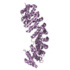
- Assembly
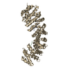
| Deposited unit | 
| ||||||||
|---|---|---|---|---|---|---|---|---|---|
| 1 |
| ||||||||
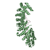
| Unit cell |
|
-Components
| #1: Protein | Mass: 49872.836 Da / Num. of mol.: 1 / Fragment: RESIDUES 72-496 Source method: isolated from a genetically manipulated source Source: (gene. exp.)  | ||
|---|---|---|---|
| #2: Protein/peptide | Mass: 1587.782 Da / Num. of mol.: 2 / Fragment: RESIDUES 839-851 Source method: isolated from a genetically manipulated source Source: (gene. exp.) #3: Water | ChemComp-HOH / |  Mass: 18.015 Da / Num. of mol.: 188 / Source method: isolated from a natural source / Formula: H2O Mass: 18.015 Da / Num. of mol.: 188 / Source method: isolated from a natural source / Formula: H2O |
-Experimental details
-Experiment
| Experiment | Method: X-RAY DIFFRACTION / Number of used crystals: 1 |
|---|
- Sample preparation
| Crystal | Density Matthews: 3.27 Å3/Da / Density % sol: 62.38 % / Description: NONE |
|---|---|
| Crystal grow | pH: 6.5 Details: 0.8 M SODIUM CITRATE, 0.1 M HEPES BUFFER (PH 6.5) AND 10 MM DTT |
-Data collection
| Diffraction | Mean temperature: 300 K |
|---|---|
| Diffraction source | Source: SYNCHROTRON / Site: Australian Synchrotron  / Beamline: MX2 / Wavelength: 0.9536 / Beamline: MX2 / Wavelength: 0.9536 |
| Detector | Date: Jul 31, 2012 |
| Radiation | Protocol: SINGLE WAVELENGTH / Monochromatic (M) / Laue (L): M / Scattering type: x-ray |
| Radiation wavelength | Wavelength: 0.9536 Å / Relative weight: 1 |
| Reflection | Resolution: 2→19.83 Å / Num. obs: 49123 / % possible obs: 99.6 % / Observed criterion σ(I): 2 / Redundancy: 7.1 % / Biso Wilson estimate: 33.84 Å2 / Rmerge(I) obs: 0.06 / Net I/σ(I): 10.3 |
| Reflection shell | Resolution: 2→2.11 Å / Redundancy: 14.55 % / Rmerge(I) obs: 0.46 / Mean I/σ(I) obs: 6.5 / % possible all: 99.3 |
- Processing
| Software |
| |||||||||||||||||||||||||||||||||||||||||||||||||||||||||||||||||||||||||||||||||||||||||||||||||||||||||||||||||||||||||||||
|---|---|---|---|---|---|---|---|---|---|---|---|---|---|---|---|---|---|---|---|---|---|---|---|---|---|---|---|---|---|---|---|---|---|---|---|---|---|---|---|---|---|---|---|---|---|---|---|---|---|---|---|---|---|---|---|---|---|---|---|---|---|---|---|---|---|---|---|---|---|---|---|---|---|---|---|---|---|---|---|---|---|---|---|---|---|---|---|---|---|---|---|---|---|---|---|---|---|---|---|---|---|---|---|---|---|---|---|---|---|---|---|---|---|---|---|---|---|---|---|---|---|---|---|---|---|---|
| Refinement | Method to determine structure: MOLECULAR REPLACEMENT Starting model: PDB ENTRY 1IAL Resolution: 2→19.83 Å / Cor.coef. Fo:Fc: 0.9521 / Cor.coef. Fo:Fc free: 0.9417 / SU R Cruickshank DPI: 0.115 / Cross valid method: THROUGHOUT / σ(F): 0 / SU R Blow DPI: 0.116 / SU Rfree Blow DPI: 0.107 / SU Rfree Cruickshank DPI: 0.107
| |||||||||||||||||||||||||||||||||||||||||||||||||||||||||||||||||||||||||||||||||||||||||||||||||||||||||||||||||||||||||||||
| Displacement parameters | Biso mean: 40.9 Å2
| |||||||||||||||||||||||||||||||||||||||||||||||||||||||||||||||||||||||||||||||||||||||||||||||||||||||||||||||||||||||||||||
| Refine analyze | Luzzati coordinate error obs: 0.25 Å | |||||||||||||||||||||||||||||||||||||||||||||||||||||||||||||||||||||||||||||||||||||||||||||||||||||||||||||||||||||||||||||
| Refinement step | Cycle: LAST / Resolution: 2→19.83 Å
| |||||||||||||||||||||||||||||||||||||||||||||||||||||||||||||||||||||||||||||||||||||||||||||||||||||||||||||||||||||||||||||
| Refine LS restraints |
| |||||||||||||||||||||||||||||||||||||||||||||||||||||||||||||||||||||||||||||||||||||||||||||||||||||||||||||||||||||||||||||
| LS refinement shell | Resolution: 2→2.05 Å / Total num. of bins used: 20
| |||||||||||||||||||||||||||||||||||||||||||||||||||||||||||||||||||||||||||||||||||||||||||||||||||||||||||||||||||||||||||||
| Refinement TLS params. | Method: refined / Refine-ID: X-RAY DIFFRACTION
| |||||||||||||||||||||||||||||||||||||||||||||||||||||||||||||||||||||||||||||||||||||||||||||||||||||||||||||||||||||||||||||
| Refinement TLS group |
|
