 Movie
Movie Controller

[English] 日本語
 Yorodumi
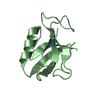
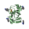
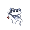
- PDB-3vj7: Crystal structure of the carboxy-terminal ribonuclease domain of ... -
+ Open data

- Basic information
| Entry | Database: PDB / ID: 3vj7 | ||||||
|---|---|---|---|---|---|---|---|
| Title | Crystal structure of the carboxy-terminal ribonuclease domain of Colicin E5 R33Q mutant | ||||||
 Components Components | Colicin-E5 | ||||||
 Keywords Keywords |  HYDROLASE / ribonuclease HYDROLASE / ribonuclease | ||||||
| Function / homology |  Function and homology information Function and homology informationRNA nuclease activity / endonuclease activity / killing of cells of another organism / Hydrolases; Acting on ester bonds / defense response to bacteriumSimilarity search - Function | ||||||
| Biological species |  Escherichia coli (E. coli) Escherichia coli (E. coli) | ||||||
| Method | X-RAY DIFFRACTION / MOLECULAR REPLACEMENT / Resolution: 2.3 Å | ||||||
 Authors Authors | Yajima, S. / Inoue, S. / Fushinobu, S. / Ogawa, T. / Hidaka, M. / Masaki, H. | ||||||
 Citation Citation | Journal: J.Biochem. / Year: 2012 Title: Identification of the catalytic residues of sequence-specific and histidine-free ribonuclease colicin E5 Authors: Inoue-Ito, S. / Yajima, S. / Fushinobu, S. / Nakamura, S. / Ogawa, T. / Hidaka, M. / Masaki, H. | ||||||
| History |
|
- Structure visualization
| Structure viewer | Molecule: MolmilJmol/JSmol |
|---|
- Downloads & links
-Download
| PDBx/mmCIF format | 3vj7.cif.gz | 35.1 KB | Display | PDBx/mmCIF format |
|---|---|---|---|---|
| PDB format | pdb3vj7.ent.gz | 22 KB | Display | PDB format |
| PDBx/mmJSON format | 3vj7.json.gz | Tree view | PDBx/mmJSON format | |
| Others |  Other downloads Other downloads |
-Validation report
| Arichive directory | https://data.pdbj.org/pub/pdb/validation_reports/vj/3vj7ftp://data.pdbj.org/pub/pdb/validation_reports/vj/3vj7 | HTTPS FTP |
|---|
-Related structure data
| Related structure data |  3ao9C  2djhS S: Starting model for refinement C: citing same article ( |
|---|---|
| Similar structure data |


-Links
 PDBj
PDBj- Assembly
| Deposited unit | 
| ||||||||
|---|---|---|---|---|---|---|---|---|---|
| 1 |
| ||||||||
| Unit cell |
|
-Components
| #1: Protein | Mass: 12693.945 Da / Num. of mol.: 1 Fragment: C-terminal ribonuclease domain, UNP residues 66-180 Mutation: R33Q Source method: isolated from a genetically manipulated source Source: (gene. exp.) Escherichia coli (E. coli) / Gene: col / Production host: Escherichia coli (E. coli)References: UniProt: P18000, Hydrolases; Acting on ester bonds |
|---|---|
| #2: Chemical | ChemComp-3PD / 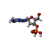  Mass: 347.221 Da / Num. of mol.: 1 / Source method: obtained synthetically / Formula: C10H14N5O7P Mass: 347.221 Da / Num. of mol.: 1 / Source method: obtained synthetically / Formula: C10H14N5O7P |
| #3: Chemical | ChemComp-UM3 / Deoxyuridine monophosphate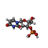  Mass: 308.182 Da / Num. of mol.: 1 / Source method: obtained synthetically / Formula: C9H13N2O8P Mass: 308.182 Da / Num. of mol.: 1 / Source method: obtained synthetically / Formula: C9H13N2O8P |
| #4: Water | ChemComp-HOH / Water Mass: 18.015 Da / Num. of mol.: 54 / Source method: isolated from a natural source / Formula: H2O Mass: 18.015 Da / Num. of mol.: 54 / Source method: isolated from a natural source / Formula: H2O |
-Experimental details
-Experiment
| Experiment | Method: X-RAY DIFFRACTION / Number of used crystals: 1 |
|---|
- Sample preparation
| Crystal | Density Matthews: 1.87 Å3/Da / Density % sol: 34.16 % |
|---|---|
| Crystal grow | Temperature: 293 K / Method: vapor diffusion, hanging drop / pH: 8 Details: 2M ammonium sulfate, 20mM TrisCl, pH 8, VAPOR DIFFUSION, HANGING DROP, temperature 293K |
-Data collection
| Diffraction | Mean temperature: 298 K |
|---|---|
| Diffraction source | Source: ROTATING ANODE / Type: MACSCIENCE / Wavelength: 1.5418 Å |
| Detector | Type: MAC Science DIP-2030 / Detector: IMAGE PLATE / Date: Nov 28, 1999 |
| Radiation | Monochromator: mirrors / Protocol: SINGLE WAVELENGTH / Monochromatic (M) / Laue (L): M / Scattering type: x-ray |
| Radiation wavelength | Wavelength: 1.5418 Å / Relative weight: 1 |
| Reflection | Resolution: 2.3→50 Å / Num. all: 4583 / Num. obs: 4574 / % possible obs: 99.8 % / Observed criterion σ(F): 2 / Observed criterion σ(I): 0 |
| Reflection shell | Resolution: 2.3→2.38 Å / % possible all: 98.8 |
- Processing
| Software |
| ||||||||||||||||||||||||||||||||||||||||||||||||||||||||||||||||||||||||||||||||||||||||||||||||||||||||||||||||||||||||||||||||||||||||||||||||||||||||||||||||||||||||||
|---|---|---|---|---|---|---|---|---|---|---|---|---|---|---|---|---|---|---|---|---|---|---|---|---|---|---|---|---|---|---|---|---|---|---|---|---|---|---|---|---|---|---|---|---|---|---|---|---|---|---|---|---|---|---|---|---|---|---|---|---|---|---|---|---|---|---|---|---|---|---|---|---|---|---|---|---|---|---|---|---|---|---|---|---|---|---|---|---|---|---|---|---|---|---|---|---|---|---|---|---|---|---|---|---|---|---|---|---|---|---|---|---|---|---|---|---|---|---|---|---|---|---|---|---|---|---|---|---|---|---|---|---|---|---|---|---|---|---|---|---|---|---|---|---|---|---|---|---|---|---|---|---|---|---|---|---|---|---|---|---|---|---|---|---|---|---|---|---|---|---|---|
| Refinement | Method to determine structure: MOLECULAR REPLACEMENT Starting model: 2DJH Resolution: 2.3→39.27 Å / Cor.coef. Fo:Fc: 0.902 / Cor.coef. Fo:Fc free: 0.875 / Cross valid method: THROUGHOUT / σ(F): 0 / ESU R Free: 0.264 / Stereochemistry target values: MAXIMUM LIKELIHOOD / Details: HYDROGENS HAVE BEEN ADDED IN THE RIDING POSITIONS
| ||||||||||||||||||||||||||||||||||||||||||||||||||||||||||||||||||||||||||||||||||||||||||||||||||||||||||||||||||||||||||||||||||||||||||||||||||||||||||||||||||||||||||
| Solvent computation | Ion probe radii: 0.8 Å / Shrinkage radii: 0.8 Å / VDW probe radii: 1.4 Å / Solvent model: MASK | ||||||||||||||||||||||||||||||||||||||||||||||||||||||||||||||||||||||||||||||||||||||||||||||||||||||||||||||||||||||||||||||||||||||||||||||||||||||||||||||||||||||||||
| Displacement parameters | Biso mean: 21.526 Å2
| ||||||||||||||||||||||||||||||||||||||||||||||||||||||||||||||||||||||||||||||||||||||||||||||||||||||||||||||||||||||||||||||||||||||||||||||||||||||||||||||||||||||||||
| Refinement step | Cycle: LAST / Resolution: 2.3→39.27 Å
| ||||||||||||||||||||||||||||||||||||||||||||||||||||||||||||||||||||||||||||||||||||||||||||||||||||||||||||||||||||||||||||||||||||||||||||||||||||||||||||||||||||||||||
| Refine LS restraints |
| ||||||||||||||||||||||||||||||||||||||||||||||||||||||||||||||||||||||||||||||||||||||||||||||||||||||||||||||||||||||||||||||||||||||||||||||||||||||||||||||||||||||||||
| LS refinement shell | Resolution: 2.301→2.36 Å / Total num. of bins used: 20
|
