 Movie
Movie Controller

[English] 日本語
 Yorodumi
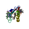
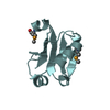
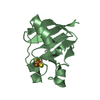
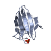
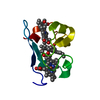
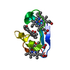
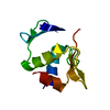
- PDB-6lk1: Ultrahigh resolution X-ray structure of Ferredoxin I from C. rein... -
+ Open data

- Basic information
| Entry | Database: PDB / ID: 6lk1 | ||||||
|---|---|---|---|---|---|---|---|
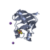
| Title | Ultrahigh resolution X-ray structure of Ferredoxin I from C. reinhardtii | ||||||
 Components Components | Ferredoxin, chloroplastic | ||||||
 Keywords Keywords | ELECTRON TRANSPORT / [2Fe2S] cluster | ||||||
| Function / homology |  Function and homology information Function and homology informationiron-sulfur cluster binding / chloroplast / electron transport chain / 2 iron, 2 sulfur cluster binding / electron transfer activity / metal ion binding Similarity search - Function | ||||||
| Biological species |   Chlamydomonas reinhardtii (plant) Chlamydomonas reinhardtii (plant) | ||||||
| Method |  X-RAY DIFFRACTION / SYNCHROTRON / MOLECULAR REPLACEMENT / Resolution: 0.9 Å X-RAY DIFFRACTION / SYNCHROTRON / MOLECULAR REPLACEMENT / Resolution: 0.9 Å | ||||||
 Authors Authors | Onishi, Y. / Kurisu, G. / Tanaka, H. | ||||||
| Funding support |  Japan, 1items Japan, 1items
| ||||||
 Citation Citation | Journal: J.Biochem. / Year: 2020 Title: X-ray dose-dependent structural changes of the [2Fe-2S] ferredoxin from Chlamydomonas reinhardtii. Authors: Ohnishi, Y. / Muraki, N. / Kiyota, D. / Okumura, H. / Baba, S. / Kawano, Y. / Kumasaka, T. / Tanaka, H. / Kurisu, G. | ||||||
| History |
|
- Structure visualization
| Structure viewer | Molecule: MolmilJmol/JSmol |
|---|
- Downloads & links
-Download
| PDBx/mmCIF format | 6lk1.cif.gz | 98.6 KB | Display | PDBx/mmCIF format |
|---|---|---|---|---|
| PDB format | pdb6lk1.ent.gz | 74.1 KB | Display | PDB format |
| PDBx/mmJSON format | 6lk1.json.gz | Tree view | PDBx/mmJSON format | |
| Others |  Other downloads Other downloads |
-Validation report
| Arichive directory | https://data.pdbj.org/pub/pdb/validation_reports/lk/6lk1ftp://data.pdbj.org/pub/pdb/validation_reports/lk/6lk1 | HTTPS FTP |
|---|
-Related structure data
| Related structure data |  6kumC 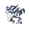 6kv0C  1awdS S: Starting model for refinement C: citing same article ( |
|---|---|
| Similar structure data |
-Links
 PDBj
PDBj


- Assembly
| Deposited unit | 
| ||||||||
|---|---|---|---|---|---|---|---|---|---|
| 1 | 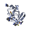
| ||||||||
| 2 | 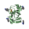
| ||||||||
| Unit cell |
|
-Components
-Protein , 1 types, 2 molecules AB
| #1: Protein | Mass: 9984.993 Da / Num. of mol.: 2 Source method: isolated from a genetically manipulated source Source: (gene. exp.) Chlamydomonas reinhardtii (plant) / Gene: PETF / Production host:  |
|---|
-Non-polymers , 5 types, 171 molecules 


| #2: Chemical |  Mass: 175.820 Da / Num. of mol.: 2 / Source method: obtained synthetically / Formula: Fe2S2 / Feature type: SUBJECT OF INVESTIGATION Mass: 175.820 Da / Num. of mol.: 2 / Source method: obtained synthetically / Formula: Fe2S2 / Feature type: SUBJECT OF INVESTIGATION#3: Chemical | ChemComp-BEN /  Mass: 120.152 Da / Num. of mol.: 10 / Source method: obtained synthetically / Formula: C7H8N2 Mass: 120.152 Da / Num. of mol.: 10 / Source method: obtained synthetically / Formula: C7H8N2#4: Chemical | ChemComp-SCN / |  Mass: 58.082 Da / Num. of mol.: 1 / Source method: obtained synthetically / Formula: CNS Mass: 58.082 Da / Num. of mol.: 1 / Source method: obtained synthetically / Formula: CNS#5: Chemical | ChemComp-SO4 / |  Mass: 96.063 Da / Num. of mol.: 1 / Source method: obtained synthetically / Formula: SO4 Mass: 96.063 Da / Num. of mol.: 1 / Source method: obtained synthetically / Formula: SO4#6: Water | ChemComp-HOH / | Mass: 18.015 Da / Num. of mol.: 157 / Source method: isolated from a natural source / Formula: H2O |
|---|
-Details
| Has ligand of interest | Y |
|---|
-Experimental details
-Experiment
| Experiment | Method: X-RAY DIFFRACTION / Number of used crystals: 1 |
|---|
- Sample preparation
| Crystal | Density Matthews: 1.91 Å3/Da / Density % sol: 35.61 % |
|---|---|
| Crystal grow | Temperature: 298 K / Method: vapor diffusion Details: Ammonium sulfate Sodium citrate Sodium thiocyanate Benzamidine Hydrochloride |
-Data collection
| Diffraction | Mean temperature: 100 K / Serial crystal experiment: N |
|---|---|
| Diffraction source | Source: SYNCHROTRON / Site: SPring-8 / Beamline: BL44XU / Wavelength: 0.9 Å |
| Detector | Type: RAYONIX MX300HE / Detector: CCD / Date: Jan 17, 2015 |
| Radiation | Protocol: SINGLE WAVELENGTH / Monochromatic (M) / Laue (L): M / Scattering type: x-ray |
| Radiation wavelength | Wavelength: 0.9 Å / Relative weight: 1 |
| Reflection | Resolution: 0.9→37.96 Å / Num. obs: 109765 / % possible obs: 99.2 % / Redundancy: 4.736 % / Rmerge(I) obs: 0.061 / Net I/σ(I): 12.9 |
| Reflection shell | Resolution: 0.9→0.92 Å / Redundancy: 3.188 % / Rmerge(I) obs: 0.729 / Mean I/σ(I) obs: 1.7 / Num. unique obs: 7995 / Rrim(I) all: 0.35 / % possible all: 97.6 |
- Processing
| Software |
| ||||||||||||||||||||
|---|---|---|---|---|---|---|---|---|---|---|---|---|---|---|---|---|---|---|---|---|---|
| Refinement | Method to determine structure: MOLECULAR REPLACEMENT Starting model: 1AWD Resolution: 0.9→37.96 Å / Cross valid method: THROUGHOUT
| ||||||||||||||||||||
| Displacement parameters | Biso max: 136.17 Å2 / Biso min: 3.93 Å2 | ||||||||||||||||||||
| Refinement step | Cycle: LAST / Resolution: 0.9→37.96 Å
|
