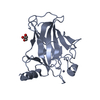
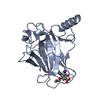
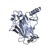
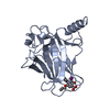
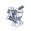
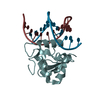
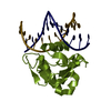
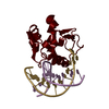
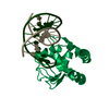
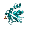
the crystallographic asymmetric unit contains two apo IBDs and four IBD-alphaSCS complexes that are arranged as a pseudocontinous helix. Because extra IBD was used in the crystallization, there are extra apo IBDs.
-
Components
#1: DNA chain
ALPHA-SCSINR
Mass: 3597.367 Da / Num. of mol.: 4 / Source method: obtained synthetically / Details: the alpha-SCS Inr promoter element, DNA site
#2: DNA chain
ALPHA-SCSINR
Mass: 3726.453 Da / Num. of mol.: 4 / Source method: obtained synthetically
#3: Protein
39kDainitiatorbindingprotein
Mass: 15344.390 Da / Num. of mol.: 6 / Fragment: residues 1-126 Source method: isolated from a genetically manipulated source Source: (gene. exp.) Trichomonas vaginalis (eukaryote) / Gene: Trichomonas vaginalis:IBP39 / Plasmid: pEQ60 / Production host: Escherichia coli (E. coli) / References: UniProt: Q95VR4
In the structure databanks used in Yorodumi, some data are registered as the other names, "COVID-19 virus" and "2019-nCoV". Here are the details of the virus and the list of structure data.
Jan 31, 2019. EMDB accession codes are about to change! (news from PDBe EMDB page)
EMDB accession codes are about to change! (news from PDBe EMDB page)
The allocation of 4 digits for EMDB accession codes will soon come to an end. Whilst these codes will remain in use, new EMDB accession codes will include an additional digit and will expand incrementally as the available range of codes is exhausted. The current 4-digit format prefixed with “EMD-” (i.e. EMD-XXXX) will advance to a 5-digit format (i.e. EMD-XXXXX), and so on. It is currently estimated that the 4-digit codes will be depleted around Spring 2019, at which point the 5-digit format will come into force.
The EM Navigator/Yorodumi systems omit the EMD- prefix.
Related info.:Q: What is EMD? / ID/Accession-code notation in Yorodumi/EM Navigator
Yorodumi is a browser for structure data from EMDB, PDB, SASBDB, etc.
This page is also the successor to EM Navigator detail page, and also detail information page/front-end page for Omokage search.
The word "yorodu" (or yorozu) is an old Japanese word meaning "ten thousand". "mi" (miru) is to see.
Related info.:EMDB / PDB / SASBDB / Comparison of 3 databanks / Yorodumi Search / Aug 31, 2016. New EM Navigator & Yorodumi / Yorodumi Papers / Jmol/JSmol / Function and homology information / Changes in new EM Navigator and Yorodumi
 Movie
Movie Controller

 Yorodumi
 Open data
 Basic information
 Components
 Keywords
 Function and homology information
 Trichomonas vaginalis (eukaryote)
 X-RAY DIFFRACTION /
  Authors
 Citation
 Structure visualization
 Downloads & links
 Other downloads
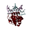


 PDBj
PDBj


 Assembly
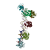


 Mass: 96.063 Da / Num. of mol.: 2 / Source method: obtained synthetically / Formula: SO4
 Sample preparation
 / Beamline: BL11-1 / Wavelength: 0.96 Å
 Processing