 Movie
Movie Controller

[English] 日本語
 Yorodumi
- PDB-1q87: Crystal structure of the C-domain of the T.vaginalis Inr binding ... -
+ Open data

- Basic information
| Entry | Database: PDB / ID: 1q87 | ||||||
|---|---|---|---|---|---|---|---|
| Title | Crystal structure of the C-domain of the T.vaginalis Inr binding protein, IBP39 (tetragonal form) | ||||||
 Components Components | 39 kDa initiator binding protein | ||||||
 Keywords Keywords | DNA BINDING PROTEIN / Initiator / Inr / Initiator binding protein / core promoter | ||||||
| Function / homology | Initiator binding domain / Initiator binding protein 39kDa, C-terminal / IBP39, C-terminal domain superfamily / Transcription-initiator DNA-binding domain IBD / Initiator binding protein 39 kDa / Winged helix DNA-binding domain superfamily / Winged helix-like DNA-binding domain superfamily / metal ion binding / 39 kDa initiator binding protein Function and homology information Function and homology information | ||||||
| Biological species |  Trichomonas vaginalis (eukaryote) Trichomonas vaginalis (eukaryote) | ||||||
| Method |  X-RAY DIFFRACTION / SYNCHROTRON / MOLECULAR REPLACEMENT / Resolution: 2.32 Å X-RAY DIFFRACTION / SYNCHROTRON / MOLECULAR REPLACEMENT / Resolution: 2.32 Å | ||||||
 Authors Authors | Schumacher, M.A. / Johnson, P.J. | ||||||
 Citation Citation | Journal: Cell(Cambridge,Mass.) / Year: 2003 Title: Structural Basis of Core Promoter Recognition in a Primitive Eukaryote Authors: Schumacher, M.A. / Lau, A.O.T. / Johnson, P.J. | ||||||
| History |
|
- Structure visualization
| Structure viewer | Molecule: MolmilJmol/JSmol |
|---|
- Downloads & links
-Download
| PDBx/mmCIF format | 1q87.cif.gz | 89.1 KB | Display | PDBx/mmCIF format |
|---|---|---|---|---|
| PDB format | pdb1q87.ent.gz | 68.1 KB | Display | PDB format |
| PDBx/mmJSON format | 1q87.json.gz | Tree view | PDBx/mmJSON format | |
| Others |  Other downloads Other downloads |
-Validation report
| Arichive directory | https://data.pdbj.org/pub/pdb/validation_reports/q8/1q87ftp://data.pdbj.org/pub/pdb/validation_reports/q8/1q87 | HTTPS FTP |
|---|
-Related structure data
| Related structure data | 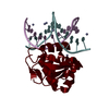 1pp7C 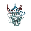 1pp8C  1q88SC 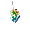 1q89C S: Starting model for refinement C: citing same article ( |
|---|---|
| Similar structure data |


-Links
 PDBj
PDBj- Assembly
| Deposited unit | 
| ||||||||
|---|---|---|---|---|---|---|---|---|---|
| 1 | 
| ||||||||
| 2 | 
| ||||||||
| Unit cell |
|
-Components
| #1: Protein | Mass: 25727.221 Da / Num. of mol.: 2 / Fragment: C-domain, residues 127-341 Source method: isolated from a genetically manipulated source Source: (gene. exp.) Trichomonas vaginalis (eukaryote) / Gene: ibp39 / Plasmid: PQE30 / Production host:  #2: Water | ChemComp-HOH / |  Mass: 18.015 Da / Num. of mol.: 61 / Source method: isolated from a natural source / Formula: H2O Mass: 18.015 Da / Num. of mol.: 61 / Source method: isolated from a natural source / Formula: H2O |
|---|
-Experimental details
-Experiment
| Experiment | Method: X-RAY DIFFRACTION / Number of used crystals: 1 |
|---|
- Sample preparation
| Crystal | Density Matthews: 2.23 Å3/Da / Density % sol: 44.97 % | ||||||||||||||||||||||||||||||
|---|---|---|---|---|---|---|---|---|---|---|---|---|---|---|---|---|---|---|---|---|---|---|---|---|---|---|---|---|---|---|---|
| Crystal grow | Temperature: 298 K / Method: vapor diffusion, hanging drop / pH: 6.5 Details: PEG 5000, ammonium sulphate, pH 6.5, VAPOR DIFFUSION, HANGING DROP, temperature 298K | ||||||||||||||||||||||||||||||
| Crystal grow | *PLUS Method: vapor diffusion | ||||||||||||||||||||||||||||||
| Components of the solutions | *PLUS
|
-Data collection
| Diffraction | Mean temperature: 100 K |
|---|---|
| Diffraction source | Source: SYNCHROTRON / Site: SSRL  / Beamline: BL9-2 / Wavelength: 1.08 Å / Beamline: BL9-2 / Wavelength: 1.08 Å |
| Detector | Type: ADSC QUANTUM 4 / Detector: CCD / Date: Nov 12, 2002 |
| Radiation | Monochromator: graphite / Protocol: SINGLE WAVELENGTH / Monochromatic (M) / Laue (L): M / Scattering type: x-ray |
| Radiation wavelength | Wavelength: 1.08 Å / Relative weight: 1 |
| Reflection | Resolution: 2.32→65 Å / Num. obs: 19164 / % possible obs: 91.6 % / Observed criterion σ(F): 11 / Observed criterion σ(I): 10.9 / Redundancy: 5.8 % / Biso Wilson estimate: 57.5 Å2 / Rmerge(I) obs: 0.042 |
| Reflection shell | Resolution: 2.32→2.38 Å / Redundancy: 4 % / Rmerge(I) obs: 0.386 / Mean I/σ(I) obs: 2 / % possible all: 91.6 |
| Reflection | *PLUS Lowest resolution: 65 Å / Num. measured all: 111463 |
- Processing
| Software |
| |||||||||||||||||||||||||
|---|---|---|---|---|---|---|---|---|---|---|---|---|---|---|---|---|---|---|---|---|---|---|---|---|---|---|
| Refinement | Method to determine structure: MOLECULAR REPLACEMENT Starting model: PDB entry 1Q88, C-domain from the C2 monoclinic form Resolution: 2.32→65 Å / Rfactor Rfree error: 0.006 / Data cutoff high absF: 1778869.52 / Data cutoff low absF: 0 / Isotropic thermal model: RESTRAINED / Cross valid method: THROUGHOUT / σ(F): 0 / Stereochemistry target values: Engh & Huber
| |||||||||||||||||||||||||
| Solvent computation | Solvent model: FLAT MODEL / Bsol: 59.8645 Å2 / ksol: 0.351692 e/Å3 | |||||||||||||||||||||||||
| Displacement parameters | Biso mean: 66.4 Å2
| |||||||||||||||||||||||||
| Refine analyze |
| |||||||||||||||||||||||||
| Refinement step | Cycle: LAST / Resolution: 2.32→65 Å
| |||||||||||||||||||||||||
| Refine LS restraints |
| |||||||||||||||||||||||||
| LS refinement shell | Resolution: 2.32→2.47 Å / Rfactor Rfree error: 0.021 / Total num. of bins used: 6
| |||||||||||||||||||||||||
| Xplor file |
| |||||||||||||||||||||||||
| Refinement | *PLUS % reflection Rfree: 5 % / Rfactor Rfree: 0.275 | |||||||||||||||||||||||||
| Solvent computation | *PLUS | |||||||||||||||||||||||||
| Displacement parameters | *PLUS | |||||||||||||||||||||||||
| Refine LS restraints | *PLUS
|
