 Movie
Movie Controller


+ Open data

- Basic information
| Entry | Database: PDB / ID: 4oku | ||||||
|---|---|---|---|---|---|---|---|
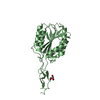
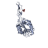
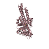
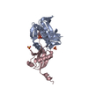
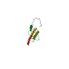
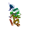
| Title | Structure of Toxoplasma gondii proMIC2 | ||||||
 Components Components | Micronemal protein MIC2 | ||||||
 Keywords Keywords | CELL ADHESION / VWA / integrin I domain / TSR domain / Adhesin / gliding motility | ||||||
| Function / homology |  Function and homology information Function and homology informationadhesion of symbiont to host cell / symbiont entry into host cell / extracellular region Similarity search - Function | ||||||
| Biological species |  | ||||||
| Method |  X-RAY DIFFRACTION / SYNCHROTRON / MOLECULAR REPLACEMENT / Resolution: 3.2 Å X-RAY DIFFRACTION / SYNCHROTRON / MOLECULAR REPLACEMENT / Resolution: 3.2 Å | ||||||
 Authors Authors | Song, G. / Springer, T.A. | ||||||
 Citation Citation | Journal: Proc.Natl.Acad.Sci.USA / Year: 2014 Title: Structures of the Toxoplasma gliding motility adhesin. Authors: Song, G. / Springer, T.A. | ||||||
| History |
|
- Structure visualization
| Structure viewer | Molecule: MolmilJmol/JSmol |
|---|
- Downloads & links
-Download
| PDBx/mmCIF format | 4oku.cif.gz | 197.8 KB | Display | PDBx/mmCIF format |
|---|---|---|---|---|
| PDB format | pdb4oku.ent.gz | 159.7 KB | Display | PDB format |
| PDBx/mmJSON format | 4oku.json.gz | Tree view | PDBx/mmJSON format | |
| Others |  Other downloads Other downloads |
-Validation report
| Arichive directory | https://data.pdbj.org/pub/pdb/validation_reports/ok/4okuftp://data.pdbj.org/pub/pdb/validation_reports/ok/4oku | HTTPS FTP |
|---|
-Related structure data


-Links
 PDBj
PDBj


- Assembly
| Deposited unit | 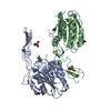
| |||||||||||||||
|---|---|---|---|---|---|---|---|---|---|---|---|---|---|---|---|---|
| 1 | 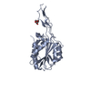
| |||||||||||||||
| 2 | 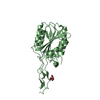
| |||||||||||||||
| 3 | 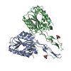
| |||||||||||||||
| Unit cell |
| |||||||||||||||
| Noncrystallographic symmetry (NCS) | NCS domain:
|
-Components
| #1: Protein | Mass: 33849.133 Da / Num. of mol.: 2 / Fragment: proMIC2 (unp residues 30-337) / Mutation: S158A Source method: isolated from a genetically manipulated source Source: (gene. exp.)  homo sapiens (human) / References: UniProt: O00816 homo sapiens (human) / References: UniProt: O00816#2: Sugar |   Type: D-saccharide, alpha linking / Mass: 180.156 Da / Num. of mol.: 2 Type: D-saccharide, alpha linking / Mass: 180.156 Da / Num. of mol.: 2Source method: isolated from a genetically manipulated source Formula: C6H12O6 Has protein modification | Y | |
|---|
-Experimental details
-Experiment
| Experiment | Method: X-RAY DIFFRACTION / Number of used crystals: 1 |
|---|
- Sample preparation
| Crystal | Density Matthews: 2.12 Å3/Da / Density % sol: 42.11 % |
|---|---|
| Crystal grow | Temperature: 293 K / Method: vapor diffusion, hanging drop / pH: 3.7 Details: 0.2 M (NH4)2SO4 and 20% PEG 3350, pH 3.7, VAPOR DIFFUSION, HANGING DROP, temperature 293K |
-Data collection
| Diffraction | Mean temperature: 100 K |
|---|---|
| Diffraction source | Source: SYNCHROTRON / Site: APS  / Beamline: 24-ID-C / Wavelength: 1 Å / Beamline: 24-ID-C / Wavelength: 1 Å |
| Detector | Type: MAR scanner 300 mm plate / Detector: IMAGE PLATE / Date: Jun 29, 2013 |
| Radiation | Monochromator: Si (111) / Protocol: SINGLE WAVELENGTH / Monochromatic (M) / Laue (L): M / Scattering type: x-ray |
| Radiation wavelength | Wavelength: 1 Å / Relative weight: 1 |
| Reflection | Resolution: 3.2→45.4 Å / Num. all: 10125 / Num. obs: 8867 / % possible obs: 87.5 % / Observed criterion σ(F): 1 / Observed criterion σ(I): 1 |
| Reflection shell | Resolution: 3.2→3.26 Å / % possible all: 65.8 |
- Processing
| Software |
| ||||||||||||||||||||||||||||||||||||||||
|---|---|---|---|---|---|---|---|---|---|---|---|---|---|---|---|---|---|---|---|---|---|---|---|---|---|---|---|---|---|---|---|---|---|---|---|---|---|---|---|---|---|
| Refinement | Method to determine structure: MOLECULAR REPLACEMENT / Resolution: 3.2→45.39 Å / SU ML: 0.49 / σ(F): 0.09 / Phase error: 39.91 / Stereochemistry target values: ML
| ||||||||||||||||||||||||||||||||||||||||
| Solvent computation | Shrinkage radii: 0.9 Å / VDW probe radii: 1.11 Å / Solvent model: FLAT BULK SOLVENT MODEL | ||||||||||||||||||||||||||||||||||||||||
| Refinement step | Cycle: LAST / Resolution: 3.2→45.39 Å
| ||||||||||||||||||||||||||||||||||||||||
| Refine LS restraints |
| ||||||||||||||||||||||||||||||||||||||||
| LS refinement shell |
| ||||||||||||||||||||||||||||||||||||||||
| Refinement TLS params. | Method: refined / Origin x: -13.1906 Å / Origin y: 38.8039 Å / Origin z: -10.8478 Å
| ||||||||||||||||||||||||||||||||||||||||
| Refinement TLS group | Selection details: all |
