 Movie
Movie Controller

[English] 日本語
 Yorodumi
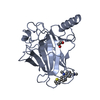
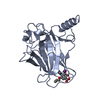
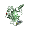
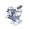
- PDB-6gge: p53 cancer mutant Y220C in complex with small-molecule stabilizer... -
+ Open data

- Basic information
| Entry | Database: PDB / ID: 6gge | ||||||
|---|---|---|---|---|---|---|---|
| Title | p53 cancer mutant Y220C in complex with small-molecule stabilizer PK9327 | ||||||
 Components Components | Cellular tumor antigen p53 | ||||||
 Keywords Keywords | DNA BINDING PROTEIN / p53 / transcription factor / tumor supressor / cancer therapy / oncogenic mutant / protein misfolding / small-molecule stabilizer / molecular chaperone | ||||||
| Function / homology |  Function and homology information Function and homology informationnegative regulation of helicase activity / Loss of function of TP53 in cancer due to loss of tetramerization ability / Regulation of TP53 Expression / signal transduction by p53 class mediator / negative regulation of G1 to G0 transition / negative regulation of glucose catabolic process to lactate via pyruvate / Transcriptional activation of cell cycle inhibitor p21 / regulation of intrinsic apoptotic signaling pathway by p53 class mediator / negative regulation of pentose-phosphate shunt / Activation of NOXA and translocation to mitochondria ...negative regulation of helicase activity / Loss of function of TP53 in cancer due to loss of tetramerization ability / Regulation of TP53 Expression / signal transduction by p53 class mediator / negative regulation of G1 to G0 transition / negative regulation of glucose catabolic process to lactate via pyruvate / Transcriptional activation of cell cycle inhibitor p21 / regulation of intrinsic apoptotic signaling pathway by p53 class mediator / negative regulation of pentose-phosphate shunt / Activation of NOXA and translocation to mitochondria / ATP-dependent DNA/DNA annealing activity / regulation of cell cycle G2/M phase transition / oligodendrocyte apoptotic process / negative regulation of miRNA processing / intrinsic apoptotic signaling pathway in response to hypoxia / oxidative stress-induced premature senescence / regulation of tissue remodeling / positive regulation of thymocyte apoptotic process / positive regulation of mitochondrial membrane permeability / germ cell nucleus / regulation of fibroblast apoptotic process / bone marrow development / circadian behavior / cellular response to actinomycin D / histone deacetylase regulator activity / positive regulation of programmed necrotic cell death / : / regulation of mitochondrial membrane permeability involved in apoptotic process / RUNX3 regulates CDKN1A transcription / T cell proliferation involved in immune response / TP53 Regulates Transcription of Death Receptors and Ligands / Activation of PUMA and translocation to mitochondria / TP53 regulates transcription of additional cell cycle genes whose exact role in the p53 pathway remain uncertain / mRNA transcription / negative regulation of glial cell proliferation / regulation of DNA damage response, signal transduction by p53 class mediator / Regulation of TP53 Activity through Association with Co-factors / negative regulation of neuroblast proliferation / Formation of Senescence-Associated Heterochromatin Foci (SAHF) / mitochondrial DNA repair / T cell lineage commitment / thymocyte apoptotic process / ER overload response / TP53 Regulates Transcription of Caspase Activators and Caspases / cardiac septum morphogenesis / B cell lineage commitment / entrainment of circadian clock by photoperiod / negative regulation of DNA replication / Zygotic genome activation (ZGA) / negative regulation of mitophagy / TP53 Regulates Transcription of Genes Involved in Cytochrome C Release / necroptotic process / PI5P Regulates TP53 Acetylation / Association of TriC/CCT with target proteins during biosynthesis / positive regulation of release of cytochrome c from mitochondria / negative regulation of telomere maintenance via telomerase / SUMOylation of transcription factors / TP53 regulates transcription of several additional cell death genes whose specific roles in p53-dependent apoptosis remain uncertain / rRNA transcription / negative regulation of reactive oxygen species metabolic process / TFIID-class transcription factor complex binding / intrinsic apoptotic signaling pathway by p53 class mediator / Transcriptional Regulation by VENTX / cellular response to UV-C / viral process / neuroblast proliferation / intrinsic apoptotic signaling pathway in response to endoplasmic reticulum stress / replicative senescence / Pyroptosis / positive regulation of RNA polymerase II transcription preinitiation complex assembly / intrinsic apoptotic signaling pathway in response to DNA damage by p53 class mediator / general transcription initiation factor binding / chromosome organization / positive regulation of execution phase of apoptosis / hematopoietic stem cell differentiation / type II interferon-mediated signaling pathway / embryonic organ development / TP53 Regulates Transcription of Genes Involved in G1 Cell Cycle Arrest / response to X-ray / hematopoietic progenitor cell differentiation / somitogenesis / negative regulation of stem cell proliferation / core promoter sequence-specific DNA binding / glial cell proliferation / cellular response to glucose starvation / negative regulation of fibroblast proliferation / cis-regulatory region sequence-specific DNA binding / mitophagy / Regulation of TP53 Activity through Acetylation / negative regulation of proteolysis / mitotic G1 DNA damage checkpoint signaling / positive regulation of intrinsic apoptotic signaling pathway / cardiac muscle cell apoptotic process / response to salt stress / transcription repressor complex / 14-3-3 protein binding / gastrulation / positive regulation of cardiac muscle cell apoptotic process / transforming growth factor beta receptor signaling pathway / reactive oxygen species metabolic process Similarity search - Function | ||||||
| Biological species |  Homo sapiens (human) Homo sapiens (human) | ||||||
| Method |  X-RAY DIFFRACTION / SYNCHROTRON / FOURIER SYNTHESIS / Resolution: 1.25000506704 Å X-RAY DIFFRACTION / SYNCHROTRON / FOURIER SYNTHESIS / Resolution: 1.25000506704 Å | ||||||
 Authors Authors | Joerger, A.C. / Bauer, M.R. | ||||||
| Funding support |  Germany, 1items Germany, 1items
| ||||||
 Citation Citation | Journal: Future Med Chem / Year: 2019 Title: A structure-guided molecular chaperone approach for restoring the transcriptional activity of the p53 cancer mutant Y220C. Authors: Bauer, M.R. / Jones, R.N. / Tareque, R.K. / Springett, B. / Dingler, F.A. / Verduci, L. / Patel, K.J. / Fersht, A.R. / Joerger, A.C. / Spencer, J. #1: Journal: Proc. Natl. Acad. Sci. U.S.A. / Year: 2006Title: Structural basis for understanding oncogenic p53 mutations and designing rescue drugs. Authors: Joerger, A.C. / Ang, H.C. / Fersht, A.R. | ||||||
| History |
|
- Structure visualization
| Structure viewer | Molecule: MolmilJmol/JSmol |
|---|
- Downloads & links
-Download
| PDBx/mmCIF format | 6gge.cif.gz | 214.7 KB | Display | PDBx/mmCIF format |
|---|---|---|---|---|
| PDB format | pdb6gge.ent.gz | 142.4 KB | Display | PDB format |
| PDBx/mmJSON format | 6gge.json.gz | Tree view | PDBx/mmJSON format | |
| Others |  Other downloads Other downloads |
-Validation report
| Arichive directory | https://data.pdbj.org/pub/pdb/validation_reports/gg/6ggeftp://data.pdbj.org/pub/pdb/validation_reports/gg/6gge | HTTPS FTP |
|---|
-Related structure data
| Related structure data |  6ggaC  6ggbC  6ggcC  6ggdC  6ggfC  2j1xS S: Starting model for refinement C: citing same article ( |
|---|---|
| Similar structure data |


-Links
 PDBj
PDBj


- Assembly
| Deposited unit | 
| ||||||||||
|---|---|---|---|---|---|---|---|---|---|---|---|
| 1 | 
| ||||||||||
| 2 | 
| ||||||||||
| Unit cell |
|
-Components
| #1: Protein | Mass: 24530.811 Da / Num. of mol.: 2 / Mutation: M133L, V203A, Y220S,N239Y, N268D Source method: isolated from a genetically manipulated source Source: (gene. exp.) Homo sapiens (human) / Gene: TP53, P53 / Production host:  #2: Chemical |   Mass: 65.409 Da / Num. of mol.: 2 / Source method: obtained synthetically / Formula: Zn Mass: 65.409 Da / Num. of mol.: 2 / Source method: obtained synthetically / Formula: Zn#3: Chemical |   Mass: 335.486 Da / Num. of mol.: 2 / Source method: obtained synthetically / Formula: C21H23N2S Mass: 335.486 Da / Num. of mol.: 2 / Source method: obtained synthetically / Formula: C21H23N2S#4: Water | ChemComp-HOH / |  Mass: 18.015 Da / Num. of mol.: 482 / Source method: isolated from a natural source / Formula: H2O Mass: 18.015 Da / Num. of mol.: 482 / Source method: isolated from a natural source / Formula: H2O |
|---|
-Experimental details
-Experiment
| Experiment | Method: X-RAY DIFFRACTION / Number of used crystals: 1 |
|---|
- Sample preparation
| Crystal | Density Matthews: 2.5 Å3/Da / Density % sol: 51 % |
|---|---|
| Crystal grow | Temperature: 293 K / Method: vapor diffusion, sitting drop Details: Protein solution: 6 mg/ml protein in 25 mM sodium phosphate, ph 7.2, 150 mm KCl, 5 mm DTT. Reservoir buffer: 100 mm HEPES, pH 7.2, 19% (w/v) polyethylene glycol 4000, 5 mm DTT. Soaking ...Details: Protein solution: 6 mg/ml protein in 25 mM sodium phosphate, ph 7.2, 150 mm KCl, 5 mm DTT. Reservoir buffer: 100 mm HEPES, pH 7.2, 19% (w/v) polyethylene glycol 4000, 5 mm DTT. Soaking buffer: Saturated solution of compound in 100 mm HEPES, ph 7.2, 10 mM sodium phosphate, ph 7.2, 19% (w/v) polyethylene glycol 4000, 20 % (v/v) glycerol, 150 mm KCl. |
-Data collection
| Diffraction | Mean temperature: 100 K |
|---|---|
| Diffraction source | Source: SYNCHROTRON / Site: Diamond  / Beamline: I03 / Wavelength: 0.98 Å / Beamline: I03 / Wavelength: 0.98 Å |
| Detector | Type: DECTRIS PILATUS3 6M / Detector: PIXEL / Date: Aug 8, 2017 |
| Radiation | Protocol: SINGLE WAVELENGTH / Monochromatic (M) / Laue (L): M / Scattering type: x-ray |
| Radiation wavelength | Wavelength: 0.98 Å / Relative weight: 1 |
| Reflection | Resolution: 1.25→29.6 Å / Num. obs: 129162 / % possible obs: 95.5 % / Redundancy: 5.3 % / Biso Wilson estimate: 13.1257049977 Å2 / Rmerge(I) obs: 0.054 / Net I/σ(I): 13.7 |
| Reflection shell | Resolution: 1.25→1.32 Å / Redundancy: 5.4 % / Rmerge(I) obs: 0.58 / Mean I/σ(I) obs: 2.8 / % possible all: 93 |
- Processing
| Software |
| |||||||||||||||||||||||||||||||||||||||||||||||||||||||||||||||||||||||||||||||||||||||||||||||||||||||||||||||||||||||||||||||||||||||||||||||||||||||||||||||||||||||||||||||||||||||||||||||||||||||||||||||||||||||||
|---|---|---|---|---|---|---|---|---|---|---|---|---|---|---|---|---|---|---|---|---|---|---|---|---|---|---|---|---|---|---|---|---|---|---|---|---|---|---|---|---|---|---|---|---|---|---|---|---|---|---|---|---|---|---|---|---|---|---|---|---|---|---|---|---|---|---|---|---|---|---|---|---|---|---|---|---|---|---|---|---|---|---|---|---|---|---|---|---|---|---|---|---|---|---|---|---|---|---|---|---|---|---|---|---|---|---|---|---|---|---|---|---|---|---|---|---|---|---|---|---|---|---|---|---|---|---|---|---|---|---|---|---|---|---|---|---|---|---|---|---|---|---|---|---|---|---|---|---|---|---|---|---|---|---|---|---|---|---|---|---|---|---|---|---|---|---|---|---|---|---|---|---|---|---|---|---|---|---|---|---|---|---|---|---|---|---|---|---|---|---|---|---|---|---|---|---|---|---|---|---|---|---|---|---|---|---|---|---|---|---|---|---|---|---|---|---|---|---|
| Refinement | Method to determine structure: FOURIER SYNTHESIS Starting model: 2J1X Resolution: 1.25000506704→29.4942478596 Å / SU ML: 0.101862459892 / Cross valid method: THROUGHOUT / σ(F): 1.32651126839 / Phase error: 16.5876228601
| |||||||||||||||||||||||||||||||||||||||||||||||||||||||||||||||||||||||||||||||||||||||||||||||||||||||||||||||||||||||||||||||||||||||||||||||||||||||||||||||||||||||||||||||||||||||||||||||||||||||||||||||||||||||||
| Solvent computation | Shrinkage radii: 0.9 Å / VDW probe radii: 1.11 Å | |||||||||||||||||||||||||||||||||||||||||||||||||||||||||||||||||||||||||||||||||||||||||||||||||||||||||||||||||||||||||||||||||||||||||||||||||||||||||||||||||||||||||||||||||||||||||||||||||||||||||||||||||||||||||
| Displacement parameters | Biso mean: 19.8700348951 Å2 | |||||||||||||||||||||||||||||||||||||||||||||||||||||||||||||||||||||||||||||||||||||||||||||||||||||||||||||||||||||||||||||||||||||||||||||||||||||||||||||||||||||||||||||||||||||||||||||||||||||||||||||||||||||||||
| Refinement step | Cycle: LAST / Resolution: 1.25000506704→29.4942478596 Å
| |||||||||||||||||||||||||||||||||||||||||||||||||||||||||||||||||||||||||||||||||||||||||||||||||||||||||||||||||||||||||||||||||||||||||||||||||||||||||||||||||||||||||||||||||||||||||||||||||||||||||||||||||||||||||
| Refine LS restraints |
| |||||||||||||||||||||||||||||||||||||||||||||||||||||||||||||||||||||||||||||||||||||||||||||||||||||||||||||||||||||||||||||||||||||||||||||||||||||||||||||||||||||||||||||||||||||||||||||||||||||||||||||||||||||||||
| LS refinement shell |
|
