 Movie
Movie Controller


+ Open data

- Basic information
| Entry | Database: PDB / ID: 3teq | ||||||
|---|---|---|---|---|---|---|---|
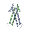
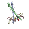
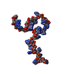
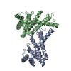
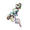
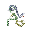
| Title | Crystal structure of SOAR domain | ||||||
 Components Components | Stromal interaction molecule 1 STIM1 STIM1 | ||||||
 Keywords Keywords | SIGNALING PROTEIN | ||||||
| Function / homology |  Function and homology information Function and homology informationstore-operated calcium entry / activation of store-operated calcium channel activity / regulation of store-operated calcium entry / cortical endoplasmic reticulum / enamel mineralization / Elevation of cytosolic Ca2+ levels / positive regulation of adenylate cyclase activity / microtubule plus-end binding / plasma membrane raft / calcium channel regulator activity ...store-operated calcium entry / activation of store-operated calcium channel activity / regulation of store-operated calcium entry / cortical endoplasmic reticulum / enamel mineralization / Elevation of cytosolic Ca2+ levels / positive regulation of adenylate cyclase activity / microtubule plus-end binding / plasma membrane raft / calcium channel regulator activity / regulation of calcium ion transport / detection of calcium ion / Ion homeostasis / sarcoplasmic reticulum membrane / Antigen activates B Cell Receptor (BCR) leading to generation of second messengers / intracellular calcium ion homeostasis / positive regulation of angiogenesis / protease binding / microtubule / calcium ion binding / endoplasmic reticulum membrane / endoplasmic reticulum / identical protein binding / plasma membraneSimilarity search - Function | ||||||
| Biological species |  Homo sapiens (human) Homo sapiens (human) | ||||||
| Method | X-RAY DIFFRACTION / SYNCHROTRON / SAD / Resolution: 1.9 Å | ||||||
 Authors Authors | Yang, X. / Jin, H. / Cai, X. / Shen, Y. | ||||||
 Citation Citation | Journal: Proc.Natl.Acad.Sci.USA / Year: 2012 Title: Structural and mechanistic insights into the activation of Stromal interaction molecule 1 (STIM1). Authors: Yang, X. / Jin, H. / Cai, X. / Li, S. / Shen, Y. | ||||||
| History |
|
- Structure visualization
| Structure viewer | Molecule: MolmilJmol/JSmol |
|---|
- Downloads & links
-Download
| PDBx/mmCIF format | 3teq.cif.gz | 105.1 KB | Display | PDBx/mmCIF format |
|---|---|---|---|---|
| PDB format | pdb3teq.ent.gz | 82.1 KB | Display | PDB format |
| PDBx/mmJSON format | 3teq.json.gz | Tree view | PDBx/mmJSON format | |
| Others |  Other downloads Other downloads |
-Validation report
| Arichive directory | https://data.pdbj.org/pub/pdb/validation_reports/te/3teqftp://data.pdbj.org/pub/pdb/validation_reports/te/3teq | HTTPS FTP |
|---|
-Related structure data

-Links
 PDBj
PDBj


- Assembly
| Deposited unit | 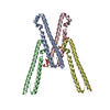
| ||||||||
|---|---|---|---|---|---|---|---|---|---|
| 1 | 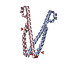
| ||||||||
| 2 | 
| ||||||||
| 3 | 
| ||||||||
| Unit cell |
|
-Components
| #1: Protein | STIM1 Mass: 11713.393 Da / Num. of mol.: 4 / Fragment: SOAR domain (UNP RESIDUES 344-444) / Mutation: L374M, V419A, C437T Source method: isolated from a genetically manipulated source Source: (gene. exp.) Homo sapiens (human) / Gene: STIM1, GOK / Production host:  Escherichia coli (E. coli) / References: UniProt: Q13586 Escherichia coli (E. coli) / References: UniProt: Q13586#2: Chemical | ChemComp-PO4 / Phosphate  Mass: 94.971 Da / Num. of mol.: 5 / Source method: obtained synthetically / Formula: PO4 Mass: 94.971 Da / Num. of mol.: 5 / Source method: obtained synthetically / Formula: PO4#3: Water | ChemComp-HOH / | Water Mass: 18.015 Da / Num. of mol.: 583 / Source method: isolated from a natural source / Formula: H2O Mass: 18.015 Da / Num. of mol.: 583 / Source method: isolated from a natural source / Formula: H2O |
|---|
-Experimental details
-Experiment
| Experiment | Method: X-RAY DIFFRACTION / Number of used crystals: 2 |
|---|
- Sample preparation
| Crystal | Density Matthews: 2.3 Å3/Da / Density % sol: 46.45 % |
|---|---|
| Crystal grow | Temperature: 277 K / Method: vapor diffusion, sitting drop / pH: 6.5 Details: 0.1M Bis-Tris pH 6.5, 10% PEG3350, 0.2M Ammonium dibasic phosphate, VAPOR DIFFUSION, SITTING DROP, temperature 277K |
-Data collection
| Diffraction |
| ||||||||||||||||||
|---|---|---|---|---|---|---|---|---|---|---|---|---|---|---|---|---|---|---|---|
| Diffraction source |
| ||||||||||||||||||
| Detector |
| ||||||||||||||||||
| Radiation |
| ||||||||||||||||||
| Radiation wavelength | Wavelength: 0.9792 Å / Relative weight: 1 | ||||||||||||||||||
| Reflection | Resolution: 1.8→50 Å / Num. all: 40114 / Num. obs: 36416 / % possible obs: 90.8 % / Observed criterion σ(F): 2 / Observed criterion σ(I): 2 / Redundancy: 13.5 % / Biso Wilson estimate: 19.2 Å2 / Rsym value: 0.079 / Net I/σ(I): 2.9 | ||||||||||||||||||
| Reflection shell | Resolution: 1.8→1.86 Å / % possible all: 66.3 |

- Processing
| Software |
| ||||||||||||||||||||||||||||||||||||
|---|---|---|---|---|---|---|---|---|---|---|---|---|---|---|---|---|---|---|---|---|---|---|---|---|---|---|---|---|---|---|---|---|---|---|---|---|---|
| Refinement | Method to determine structure: SAD / Resolution: 1.9→25.2 Å / Rfactor Rfree error: 0.006 / Data cutoff high absF: 2320392.11 / Data cutoff low absF: 0 / Isotropic thermal model: RESTRAINED / Cross valid method: THROUGHOUT / σ(F): 0 / Stereochemistry target values: Engh & Huber / Details: BULK SOLVENT MODEL USED
| ||||||||||||||||||||||||||||||||||||
| Solvent computation | Solvent model: FLAT MODEL / Bsol: 57.9755 Å2 / ksol: 0.4 e/Å3 | ||||||||||||||||||||||||||||||||||||
| Displacement parameters | Biso mean: 28.5 Å2
| ||||||||||||||||||||||||||||||||||||
| Refine analyze |
| ||||||||||||||||||||||||||||||||||||
| Refinement step | Cycle: LAST / Resolution: 1.9→25.2 Å
| ||||||||||||||||||||||||||||||||||||
| Refine LS restraints |
| ||||||||||||||||||||||||||||||||||||
| Refine LS restraints NCS | NCS model details: CONSTR | ||||||||||||||||||||||||||||||||||||
| LS refinement shell | Resolution: 1.9→2.02 Å / Rfactor Rfree error: 0.02 / Total num. of bins used: 6
| ||||||||||||||||||||||||||||||||||||
| Xplor file |
|
