 Movie
Movie Controller

[English] 日本語
 Yorodumi
- PDB-3q2p: Reduced sweetness of a monellin (MNEI) mutant results from increa... -
+ Open data

- Basic information
| Entry | Database: PDB / ID: 3q2p | ||||||
|---|---|---|---|---|---|---|---|
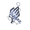
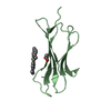
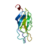
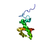
| Title | Reduced sweetness of a monellin (MNEI) mutant results from increased protein flexibility and disruption of a distant poly-(L-proline) II helix | ||||||
 Components Components | Monellin chain B/Monellin chain A chimeric protein | ||||||
 Keywords Keywords | PLANT PROTEIN / sweet protein / sweet receptor / T1R2:T1R3 | ||||||
| Function / homology |  Function and homology information Function and homology informationMonellin, A chain / Monellin, A chain superfamily / Monellin, B chain / : / Monellin / Monellin / Nuclear Transport Factor 2; Chain: A, - #10 / Cystatin superfamily / Nuclear Transport Factor 2; Chain: A, / Roll / Alpha Beta Similarity search - Domain/homology | ||||||
| Biological species |  Dioscoreophyllum cumminsii (serendipity berry) Dioscoreophyllum cumminsii (serendipity berry) | ||||||
| Method |  X-RAY DIFFRACTION / MOLECULAR REPLACEMENT / Resolution: 2.341 Å X-RAY DIFFRACTION / MOLECULAR REPLACEMENT / Resolution: 2.341 Å | ||||||
 Authors Authors | Templeton, C.M. / Hobbs, J.R. / Munger, S.D. / Conn, G.L. | ||||||
 Citation Citation | Journal: Chem Senses / Year: 2011 Title: Reduced Sweetness of a Monellin (MNEI) Mutant Results from Increased Protein Flexibility and Disruption of a Distant Poly-(L-Proline) II Helix. Authors: Templeton, C.M. / Ostovar Pour, S. / Hobbs, J.R. / Blanch, E.W. / Munger, S.D. / Conn, G.L. | ||||||
| History |
|
- Structure visualization
| Structure viewer | Molecule: MolmilJmol/JSmol |
|---|
- Downloads & links
-Download
| PDBx/mmCIF format | 3q2p.cif.gz | 93.7 KB | Display | PDBx/mmCIF format |
|---|---|---|---|---|
| PDB format | pdb3q2p.ent.gz | 72.5 KB | Display | PDB format |
| PDBx/mmJSON format | 3q2p.json.gz | Tree view | PDBx/mmJSON format | |
| Others |  Other downloads Other downloads |
-Validation report
| Summary document | 3q2p_validation.pdf.gz | 448.6 KB | Display | wwPDB validaton report |
|---|---|---|---|---|
| Full document | 3q2p_full_validation.pdf.gz | 453 KB | Display | |
| Data in XML | 3q2p_validation.xml.gz | 18.1 KB | Display | |
| Data in CIF | 3q2p_validation.cif.gz | 25.5 KB | Display | |
| Arichive directory | https://data.pdbj.org/pub/pdb/validation_reports/q2/3q2pftp://data.pdbj.org/pub/pdb/validation_reports/q2/3q2p | HTTPS FTP |
-Related structure data
| Related structure data |  3pxmC  3pyjC  2o9uS C: citing same article ( S: Starting model for refinement |
|---|---|
| Similar structure data |

-Links
 PDBj
PDBj
- Assembly
| Deposited unit | 
| ||||||||
|---|---|---|---|---|---|---|---|---|---|
| 1 | 
| ||||||||
| 2 | 
| ||||||||
| 3 | 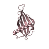
| ||||||||
| 4 | 
| ||||||||
| Unit cell |
|
-Components
| #1: Protein | Mass: 11405.021 Da / Num. of mol.: 4 / Mutation: G16A, V37A Source method: isolated from a genetically manipulated source Source: (gene. exp.) Dioscoreophyllum cumminsii (serendipity berry)Plasmid: pET22b / Production host:  #2: Chemical | ChemComp-NA / |   Mass: 22.990 Da / Num. of mol.: 1 / Source method: obtained synthetically / Formula: Na Mass: 22.990 Da / Num. of mol.: 1 / Source method: obtained synthetically / Formula: Na#3: Water | ChemComp-HOH / |  Mass: 18.015 Da / Num. of mol.: 202 / Source method: isolated from a natural source / Formula: H2O Mass: 18.015 Da / Num. of mol.: 202 / Source method: isolated from a natural source / Formula: H2O |
|---|
-Experimental details
-Experiment
| Experiment | Method: X-RAY DIFFRACTION / Number of used crystals: 1 |
|---|
- Sample preparation
| Crystal | Density Matthews: 2.27 Å3/Da / Density % sol: 45.85 % |
|---|---|
| Crystal grow | Temperature: 293.15 K / Method: vapor diffusion, hanging drop / pH: 4.2 Details: 200 mM NaCl, 100 mM phosphate-citrate, 20% w/v PEG8000, pH 4.2, VAPOR DIFFUSION, HANGING DROP, temperature 293.15K |
-Data collection
| Diffraction | Mean temperature: 200 K |
|---|---|
| Diffraction source | Source: ROTATING ANODE / Type: RIGAKU / Wavelength: 1.5418 Å |
| Detector | Type: RIGAKU / Detector: IMAGE PLATE / Date: Apr 10, 2008 |
| Radiation | Protocol: SINGLE WAVELENGTH / Monochromatic (M) / Laue (L): M / Scattering type: x-ray |
| Radiation wavelength | Wavelength: 1.5418 Å / Relative weight: 1 |
| Reflection | Resolution: 2.34→40 Å / Num. all: 16980 / Num. obs: 16708 / % possible obs: 98.4 % / Observed criterion σ(F): 0 / Observed criterion σ(I): 0 / Redundancy: 7.3 % / Rmerge(I) obs: 0.104 / Net I/σ(I): 16 |
| Reflection shell | Resolution: 2.34→2.49 Å / Redundancy: 5.5 % / Rmerge(I) obs: 0.531 / Mean I/σ(I) obs: 3.8 / Num. unique all: 2480 / % possible all: 90.8 |
- Processing
| Software |
| |||||||||||||||||||||||||||||||||||||||||||||||||
|---|---|---|---|---|---|---|---|---|---|---|---|---|---|---|---|---|---|---|---|---|---|---|---|---|---|---|---|---|---|---|---|---|---|---|---|---|---|---|---|---|---|---|---|---|---|---|---|---|---|---|
| Refinement | Method to determine structure: MOLECULAR REPLACEMENT Starting model: PDB entry 2O9U Resolution: 2.341→31.4 Å / SU ML: 0.35 / σ(F): 0 / Stereochemistry target values: ML
| |||||||||||||||||||||||||||||||||||||||||||||||||
| Solvent computation | Shrinkage radii: 0.9 Å / VDW probe radii: 1.11 Å / Solvent model: FLAT BULK SOLVENT MODEL / Bsol: 31.622 Å2 / ksol: 0.332 e/Å3 | |||||||||||||||||||||||||||||||||||||||||||||||||
| Displacement parameters |
| |||||||||||||||||||||||||||||||||||||||||||||||||
| Refinement step | Cycle: LAST / Resolution: 2.341→31.4 Å
| |||||||||||||||||||||||||||||||||||||||||||||||||
| Refine LS restraints |
| |||||||||||||||||||||||||||||||||||||||||||||||||
| LS refinement shell |
|
