 Movie
Movie Controller

[English] 日本語
 Yorodumi
- PDB-3pxc: Impact of BRCA1 BRCT domain missense substitutions on phospho-pep... -
+ Open data

- Basic information
| Entry | Database: PDB / ID: 3pxc | ||||||
|---|---|---|---|---|---|---|---|
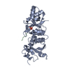
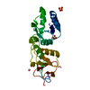
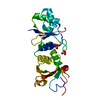
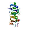
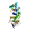
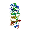
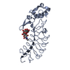
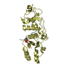
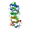
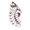
| Title | Impact of BRCA1 BRCT domain missense substitutions on phospho-peptide recognition: R1699Q | ||||||
 Components Components | Breast cancer type 1 susceptibility protein | ||||||
 Keywords Keywords |  PROTEIN BINDING / BRCA1 protein / Missense / phosphopeptide recognition / BRCT tandem repeats / BACH1 Ctip Abraxas / nuclear protein PROTEIN BINDING / BRCA1 protein / Missense / phosphopeptide recognition / BRCT tandem repeats / BACH1 Ctip Abraxas / nuclear protein | ||||||
| Function / homology |  Function and homology information Function and homology informationDefective DNA double strand break response due to BRCA1 loss of function / Defective DNA double strand break response due to BARD1 loss of function / : / BRCA1-BARD1 complex / BRCA1-C complex / BRCA1-B complex / BRCA1-A complex / random inactivation of X chromosome / negative regulation of centriole replication / negative regulation of intracellular estrogen receptor signaling pathway ...Defective DNA double strand break response due to BRCA1 loss of function / Defective DNA double strand break response due to BARD1 loss of function / : / BRCA1-BARD1 complex / BRCA1-C complex / BRCA1-B complex / BRCA1-A complex / random inactivation of X chromosome / negative regulation of centriole replication / negative regulation of intracellular estrogen receptor signaling pathway / gamma-tubulin ring complex / nuclear ubiquitin ligase complex / DNA strand resection involved in replication fork processing / chordate embryonic development / negative regulation of fatty acid biosynthetic process / cellular response to indole-3-methanol / homologous recombination / lateral element / XY body / protein K6-linked ubiquitination / regulation of DNA damage checkpoint / dosage compensation by inactivation of X chromosome / negative regulation of gene expression via chromosomal CpG island methylation / Impaired BRCA2 binding to PALB2 / : / mitotic G2/M transition checkpoint / postreplication repair / DNA repair complex / RNA polymerase binding / centrosome cycle / Defective homologous recombination repair (HRR) due to BRCA1 loss of function / Defective HDR through Homologous Recombination Repair (HRR) due to PALB2 loss of BRCA1 binding function / Defective HDR through Homologous Recombination Repair (HRR) due to PALB2 loss of BRCA2/RAD51/RAD51C binding function / Homologous DNA Pairing and Strand Exchange / Resolution of D-loop Structures through Synthesis-Dependent Strand Annealing (SDSA) / Resolution of D-loop Structures through Holliday Junction Intermediates / HDR through Single Strand Annealing (SSA) / Impaired BRCA2 binding to RAD51 / response to ionizing radiation / DNA-binding transcription activator activity / intracellular non-membrane-bounded organelle / Transcriptional Regulation by E2F6 / mitotic G2 DNA damage checkpoint signaling / Presynaptic phase of homologous DNA pairing and strand exchange / negative regulation of cell cycle / positive regulation of vascular endothelial growth factor production / negative regulation of reactive oxygen species metabolic process / localization / regulation of DNA repair / protein autoubiquitination / ubiquitin ligase complex / SUMOylation of DNA damage response and repair proteins / negative regulation of extrinsic apoptotic signaling pathway via death domain receptors / positive regulation of DNA repair / Meiotic synapsis / tubulin binding / male germ cell nucleus / chromosome segregation / cellular response to ionizing radiation / TP53 Regulates Transcription of DNA Repair Genes / Nonhomologous End-Joining (NHEJ) / double-strand break repair via homologous recombination / RING-type E3 ubiquitin transferase / HDR through Homologous Recombination (HRR) / G2/M DNA damage checkpoint / negative regulation of cell growth / Metalloprotease DUBs / Meiotic recombination / ubiquitin-protein transferase activity / fatty acid biosynthetic process / positive regulation of angiogenesis / intrinsic apoptotic signaling pathway in response to DNA damage / KEAP1-NFE2L2 pathway / double-strand break repair / p53 binding / Recruitment and ATM-mediated phosphorylation of repair and signaling proteins at DNA double strand breaks / chromosome / Neddylation / cellular response to tumor necrosis factor / Processing of DNA double-strand break ends / Regulation of TP53 Activity through Phosphorylation / damaged DNA binding / transcription coactivator activity / protein ubiquitination / nuclear body / transcription cis-regulatory region binding / regulation of cell cycle / ribonucleoprotein complex / DNA repair / negative regulation of DNA-templated transcription / DNA damage response / ubiquitin protein ligase binding / positive regulation of gene expression / regulation of transcription by RNA polymerase II / positive regulation of DNA-templated transcription / enzyme binding / positive regulation of transcription by RNA polymerase II / protein-containing complex / DNA binding / RNA bindingSimilarity search - Function | ||||||
| Biological species |  Homo sapiens (human) Homo sapiens (human) | ||||||
| Method | X-RAY DIFFRACTION / SYNCHROTRON / MOLECULAR REPLACEMENT / Resolution: 2.8 Å | ||||||
 Authors Authors | Coquelle, N. / Green, R. / Glover, J.N.M. | ||||||
 Citation Citation | Journal: Biochemistry / Year: 2011 Title: Impact of BRCA1 BRCT Domain Missense Substitutions on Phosphopeptide Recognition. Authors: Coquelle, N. / Green, R. / Glover, J.N. #1: Journal: Cancer Res. / Year: 2010 Title: Comprehensive analysis of missense variations in the BRCT domain of BRCA1 by structural and functional assays. Authors: Lee, M.S. / Green, R. / Marsillac, S.M. / Coquelle, N. / Williams, R.S. / Yeung, T. / Foo, D. / Hau, D.D. / Hui, B. / Monteiro, A.N. / Glover, J.N. #2: Journal: Nat.Struct.Mol.Biol. / Year: 2004Title: Structural basis of phosphopeptide recognition by the BRCT domain of BRCA1. Authors: Williams, R.S. / Lee, M.S. / Hau, D.D. / Glover, J.N. #3: Journal: Nat.Struct.Mol.Biol. / Year: 2004Title: Structure and mechanism of BRCA1 BRCT domain recognition of phosphorylated BACH1 with implications for cancer. Authors: Clapperton, J.A. / Manke, I.A. / Lowery, D.M. / Ho, T. / Haire, L.F. / Yaffe, M.B. / Smerdon, S.J. #4: Journal: J.Biol.Chem. / Year: 2003Title: Structural consequences of a cancer-causing BRCA1-BRCT missense mutation. Authors: Williams, R.S. / Glover, J.N. | ||||||
| History |
|
- Structure visualization
| Structure viewer | Molecule: MolmilJmol/JSmol |
|---|
- Downloads & links
-Download
| PDBx/mmCIF format | 3pxc.cif.gz | 100.4 KB | Display | PDBx/mmCIF format |
|---|---|---|---|---|
| PDB format | pdb3pxc.ent.gz | 77.2 KB | Display | PDB format |
| PDBx/mmJSON format | 3pxc.json.gz | Tree view | PDBx/mmJSON format | |
| Others |  Other downloads Other downloads |
-Validation report
| Arichive directory | https://data.pdbj.org/pub/pdb/validation_reports/px/3pxcftp://data.pdbj.org/pub/pdb/validation_reports/px/3pxc | HTTPS FTP |
|---|
-Related structure data
| Related structure data |  3pxaC  3pxbC  3pxdC  3pxeC  1n5oS S: Starting model for refinement C: citing same article ( |
|---|---|
| Similar structure data |


-Links
 PDBj
PDBj


- Assembly
| Deposited unit | 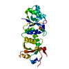
| ||||||||
|---|---|---|---|---|---|---|---|---|---|
| 1 |
| ||||||||
| Unit cell |
| ||||||||
| Components on special symmetry positions |
|
-Components
| #1: Protein | Mass: 24502.170 Da / Num. of mol.: 1 / Fragment: BRCT domain, UNP residues 1646-1859 / Mutation: R1699Q Source method: isolated from a genetically manipulated source Source: (gene. exp.) Homo sapiens (human) / Gene: BRCA1, RNF53 / Plasmid: pLM1 / Production host:  Escherichia coli (E. coli) / Strain (production host): BL21 Gold Escherichia coli (E. coli) / Strain (production host): BL21 GoldReferences: UniProt: P38398, Ligases; Forming carbon-nitrogen bonds; Acid-amino-acid ligases (peptide synthases) |
|---|---|
| #2: Chemical | ChemComp-SO4 / Sulfate  Mass: 96.063 Da / Num. of mol.: 1 / Source method: obtained synthetically / Formula: SO4 Mass: 96.063 Da / Num. of mol.: 1 / Source method: obtained synthetically / Formula: SO4 |
| #3: Chemical | ChemComp-NI / Nickel  Mass: 58.693 Da / Num. of mol.: 1 / Source method: obtained synthetically / Formula: Ni Mass: 58.693 Da / Num. of mol.: 1 / Source method: obtained synthetically / Formula: Ni |
| #4: Water | ChemComp-HOH / Water Mass: 18.015 Da / Num. of mol.: 13 / Source method: isolated from a natural source / Formula: H2O Mass: 18.015 Da / Num. of mol.: 13 / Source method: isolated from a natural source / Formula: H2O |
-Experimental details
-Experiment
| Experiment | Method: X-RAY DIFFRACTION / Number of used crystals: 1 |
|---|
- Sample preparation
| Crystal | Density Matthews: 4.73 Å3/Da / Density % sol: 73.97 % |
|---|---|
| Crystal grow | Temperature: 293 K / Method: vapor diffusion, hanging drop / pH: 8 Details: 0.8M Li2SO4 100mM Tris 5mM CaCl2 10mM NiCl2, VAPOR DIFFUSION, HANGING DROP, temperature 293K |
-Data collection
| Diffraction | Mean temperature: 100 K | |||||||||||||||||||||
|---|---|---|---|---|---|---|---|---|---|---|---|---|---|---|---|---|---|---|---|---|---|---|
| Diffraction source | Source: SYNCHROTRON / Site: ALS  / Beamline: 12.3.1 / Wavelength: 1.11584 Å / Beamline: 12.3.1 / Wavelength: 1.11584 Å | |||||||||||||||||||||
| Detector | Type: ADSC QUANTUM 315 / Detector: CCD / Date: Aug 30, 2009 | |||||||||||||||||||||
| Radiation | Monochromator: Si(111) Double crystal / Protocol: SINGLE WAVELENGTH / Monochromatic (M) / Laue (L): M / Scattering type: x-ray | |||||||||||||||||||||
| Radiation wavelength | Wavelength: 1.11584 Å / Relative weight: 1 | |||||||||||||||||||||
| Reflection | Resolution: 2.8→20 Å / Num. all: 12036 / Num. obs: 12036 / % possible obs: 98.8 % / Observed criterion σ(F): 0 / Observed criterion σ(I): 0 / Redundancy: 4.7 % / Biso Wilson estimate: 70.3 Å2 / Rsym value: 0.057 / Net I/σ(I): 16.6 | |||||||||||||||||||||
| Reflection shell |
|
- Processing
| Software |
| ||||||||||||||||||||||||||||||||||||||||||||||||||||||||||||||||||||||||||||||||||||||||||||||||||||||||||||||||||||||||||||||||||||||||||||||||||||||
|---|---|---|---|---|---|---|---|---|---|---|---|---|---|---|---|---|---|---|---|---|---|---|---|---|---|---|---|---|---|---|---|---|---|---|---|---|---|---|---|---|---|---|---|---|---|---|---|---|---|---|---|---|---|---|---|---|---|---|---|---|---|---|---|---|---|---|---|---|---|---|---|---|---|---|---|---|---|---|---|---|---|---|---|---|---|---|---|---|---|---|---|---|---|---|---|---|---|---|---|---|---|---|---|---|---|---|---|---|---|---|---|---|---|---|---|---|---|---|---|---|---|---|---|---|---|---|---|---|---|---|---|---|---|---|---|---|---|---|---|---|---|---|---|---|---|---|---|---|---|---|---|
| Refinement | Method to determine structure: MOLECULAR REPLACEMENT Starting model: 1N5O Resolution: 2.8→19.595 Å / SU ML: 0.39 / σ(F): 0 / Stereochemistry target values: Engh & Huber
| ||||||||||||||||||||||||||||||||||||||||||||||||||||||||||||||||||||||||||||||||||||||||||||||||||||||||||||||||||||||||||||||||||||||||||||||||||||||
| Solvent computation | Shrinkage radii: 0.95 Å / VDW probe radii: 1.2 Å / Solvent model: FLAT BULK SOLVENT MODEL / Bsol: 57.708 Å2 / ksol: 0.306 e/Å3 | ||||||||||||||||||||||||||||||||||||||||||||||||||||||||||||||||||||||||||||||||||||||||||||||||||||||||||||||||||||||||||||||||||||||||||||||||||||||
| Displacement parameters |
| ||||||||||||||||||||||||||||||||||||||||||||||||||||||||||||||||||||||||||||||||||||||||||||||||||||||||||||||||||||||||||||||||||||||||||||||||||||||
| Refinement step | Cycle: LAST / Resolution: 2.8→19.595 Å
| ||||||||||||||||||||||||||||||||||||||||||||||||||||||||||||||||||||||||||||||||||||||||||||||||||||||||||||||||||||||||||||||||||||||||||||||||||||||
| Refine LS restraints |
| ||||||||||||||||||||||||||||||||||||||||||||||||||||||||||||||||||||||||||||||||||||||||||||||||||||||||||||||||||||||||||||||||||||||||||||||||||||||
| LS refinement shell |
| ||||||||||||||||||||||||||||||||||||||||||||||||||||||||||||||||||||||||||||||||||||||||||||||||||||||||||||||||||||||||||||||||||||||||||||||||||||||
| Refinement TLS params. | Method: refined / Refine-ID: X-RAY DIFFRACTION
| ||||||||||||||||||||||||||||||||||||||||||||||||||||||||||||||||||||||||||||||||||||||||||||||||||||||||||||||||||||||||||||||||||||||||||||||||||||||
| Refinement TLS group |
|
