 Movie
Movie Controller

[English] 日本語
 Yorodumi
- PDB-3kaj: Apoenzyme structure of homoglutathione synthetase from Glycine ma... -
+ Open data

- Basic information
| Entry | Database: PDB / ID: 3kaj | ||||||
|---|---|---|---|---|---|---|---|
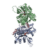
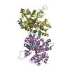
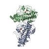
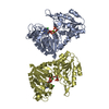
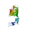
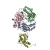
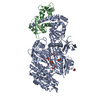
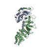
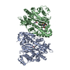
| Title | Apoenzyme structure of homoglutathione synthetase from Glycine max in open conformation | ||||||
 Components Components | Homoglutathione synthetase | ||||||
 Keywords Keywords | LIGASE / Dimer / ATP-grasp domain | ||||||
| Function / homology |  Function and homology information Function and homology informationglutathione synthase / glutathione synthase activity / glutathione binding / chloroplast / magnesium ion binding / ATP binding Similarity search - Function | ||||||
| Biological species |  | ||||||
| Method |  X-RAY DIFFRACTION / SYNCHROTRON / MOLECULAR REPLACEMENT / Resolution: 2 Å X-RAY DIFFRACTION / SYNCHROTRON / MOLECULAR REPLACEMENT / Resolution: 2 Å | ||||||
 Authors Authors | Galant, A. / Arkus, K.A.J. / Zubieta, C. / Cahoon, R.E. / Jez, J.M. | ||||||
 Citation Citation | Journal: Plant Cell / Year: 2009 Title: Structural Basis for Evolution of Product Diversity in Soybean Glutathione Biosynthesis. Authors: Galant, A. / Arkus, K.A. / Zubieta, C. / Cahoon, R.E. / Jez, J.M. | ||||||
| History |
|
- Structure visualization
| Structure viewer | Molecule: MolmilJmol/JSmol |
|---|
- Downloads & links
-Download
| PDBx/mmCIF format | 3kaj.cif.gz | 200 KB | Display | PDBx/mmCIF format |
|---|---|---|---|---|
| PDB format | pdb3kaj.ent.gz | 159.6 KB | Display | PDB format |
| PDBx/mmJSON format | 3kaj.json.gz | Tree view | PDBx/mmJSON format | |
| Others |  Other downloads Other downloads |
-Validation report
| Arichive directory | https://data.pdbj.org/pub/pdb/validation_reports/ka/3kajftp://data.pdbj.org/pub/pdb/validation_reports/ka/3kaj | HTTPS FTP |
|---|
-Related structure data
| Related structure data |  3kakC  3kalC  2hgsS C: citing same article ( S: Starting model for refinement |
|---|---|
| Similar structure data |

-Links
 PDBj
PDBj


- Assembly
| Deposited unit | 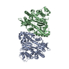
| ||||||||
|---|---|---|---|---|---|---|---|---|---|
| 1 |
| ||||||||
| Unit cell |
|
-Components
| #1: Protein | Mass: 55726.438 Da / Num. of mol.: 2 Source method: isolated from a genetically manipulated source Source: (gene. exp.)  #2: Water | ChemComp-HOH / |  Mass: 18.015 Da / Num. of mol.: 469 / Source method: isolated from a natural source / Formula: H2O Mass: 18.015 Da / Num. of mol.: 469 / Source method: isolated from a natural source / Formula: H2O |
|---|
-Experimental details
-Experiment
| Experiment | Method: X-RAY DIFFRACTION / Number of used crystals: 1 |
|---|
- Sample preparation
| Crystal | Density Matthews: 2.1 Å3/Da / Density % sol: 41.34 % |
|---|---|
| Crystal grow | Temperature: 277 K / Method: vapor diffusion, hanging drop / pH: 7 Details: 20% PEG3000, 0.1 M 3-(N-morpholino)-2-hydroxypropanesulfonic acid (MOPSO), pH 7, 0.2 M MgSO4, VAPOR DIFFUSION, HANGING DROP, temperature 277K |
-Data collection
| Diffraction | Mean temperature: 100 K |
|---|---|
| Diffraction source | Source: SYNCHROTRON / Site: SSRL  / Beamline: BL9-1 / Wavelength: 0.979 Å / Beamline: BL9-1 / Wavelength: 0.979 Å |
| Detector | Type: ADSC QUANTUM 315r / Detector: CCD / Date: Jun 18, 2007 |
| Radiation | Monochromator: Side-scattering cuberoot I-beam bent single crystal Protocol: SINGLE WAVELENGTH / Monochromatic (M) / Laue (L): M / Scattering type: x-ray |
| Radiation wavelength | Wavelength: 0.979 Å / Relative weight: 1 |
| Reflection | Resolution: 2→28.4 Å / Num. all: 62342 / Num. obs: 59099 / % possible obs: 94.8 % / Observed criterion σ(F): 0 / Observed criterion σ(I): 0 / Redundancy: 2.2 % / Biso Wilson estimate: 26 Å2 / Rsym value: 0.069 / Net I/σ(I): 13.7 |
| Reflection shell | Resolution: 2→2.05 Å / Redundancy: 2.1 % / Mean I/σ(I) obs: 3.4 / Num. unique all: 8652 / Rsym value: 0.379 / % possible all: 86.8 |
- Processing
| Software |
| ||||||||||||||||||||||||||||||||||||||||||||||||||||||||||||||||||||||||||||||||||||||||||||||||||||||||||||||||||||||||||||||||||||||||||||||||||||||||||||||||||||||||||
|---|---|---|---|---|---|---|---|---|---|---|---|---|---|---|---|---|---|---|---|---|---|---|---|---|---|---|---|---|---|---|---|---|---|---|---|---|---|---|---|---|---|---|---|---|---|---|---|---|---|---|---|---|---|---|---|---|---|---|---|---|---|---|---|---|---|---|---|---|---|---|---|---|---|---|---|---|---|---|---|---|---|---|---|---|---|---|---|---|---|---|---|---|---|---|---|---|---|---|---|---|---|---|---|---|---|---|---|---|---|---|---|---|---|---|---|---|---|---|---|---|---|---|---|---|---|---|---|---|---|---|---|---|---|---|---|---|---|---|---|---|---|---|---|---|---|---|---|---|---|---|---|---|---|---|---|---|---|---|---|---|---|---|---|---|---|---|---|---|---|---|---|
| Refinement | Method to determine structure: MOLECULAR REPLACEMENT Starting model: human glutathione synthetase, 2HGS Resolution: 2→28.38 Å / Cor.coef. Fo:Fc: 0.942 / Cor.coef. Fo:Fc free: 0.894 / SU B: 4.992 / SU ML: 0.143 / Cross valid method: THROUGHOUT / σ(F): 0 / σ(I): 0 / ESU R: 0.218 / ESU R Free: 0.201 / Stereochemistry target values: MAXIMUM LIKELIHOOD / Details: HYDROGENS HAVE BEEN ADDED IN THE RIDING POSITIONS
| ||||||||||||||||||||||||||||||||||||||||||||||||||||||||||||||||||||||||||||||||||||||||||||||||||||||||||||||||||||||||||||||||||||||||||||||||||||||||||||||||||||||||||
| Solvent computation | Ion probe radii: 0.8 Å / Shrinkage radii: 0.8 Å / VDW probe radii: 1.4 Å / Solvent model: MASK | ||||||||||||||||||||||||||||||||||||||||||||||||||||||||||||||||||||||||||||||||||||||||||||||||||||||||||||||||||||||||||||||||||||||||||||||||||||||||||||||||||||||||||
| Displacement parameters | Biso mean: 25.989 Å2
| ||||||||||||||||||||||||||||||||||||||||||||||||||||||||||||||||||||||||||||||||||||||||||||||||||||||||||||||||||||||||||||||||||||||||||||||||||||||||||||||||||||||||||
| Refinement step | Cycle: LAST / Resolution: 2→28.38 Å
| ||||||||||||||||||||||||||||||||||||||||||||||||||||||||||||||||||||||||||||||||||||||||||||||||||||||||||||||||||||||||||||||||||||||||||||||||||||||||||||||||||||||||||
| Refine LS restraints |
| ||||||||||||||||||||||||||||||||||||||||||||||||||||||||||||||||||||||||||||||||||||||||||||||||||||||||||||||||||||||||||||||||||||||||||||||||||||||||||||||||||||||||||
| LS refinement shell | Resolution: 2→2.052 Å / Total num. of bins used: 20
|
