 Movie
Movie Controller


+ Open data

- Basic information
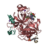
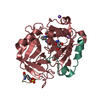
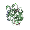
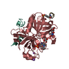
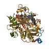
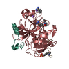
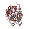
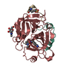
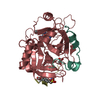
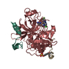
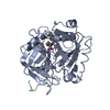
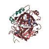
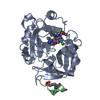
| Entry | Database: PDB / ID: 3hat | ||||||||||||
|---|---|---|---|---|---|---|---|---|---|---|---|---|---|
| Title | ACTIVE SITE MIMETIC INHIBITION OF THROMBIN | ||||||||||||
 Components Components |
| ||||||||||||
 Keywords Keywords | HYDROLASE/HYDROLASE INHIBITOR / COMPLEX (SERINE PROTEASE-INHIBITOR) / COMPLEX (SERINE PROTEASE-INHIBITOR) complex / HYDROLASE-HYDROLASE INHIBITOR COMPLEX | ||||||||||||
| Function / homology |  Function and homology information Function and homology informationpositive regulation of lipid kinase activity / positive regulation of phospholipase C-activating G protein-coupled receptor signaling pathway / cytolysis by host of symbiont cells / thrombospondin receptor activity / Defective factor XII causes hereditary angioedema /  thrombin / neutrophil-mediated killing of gram-negative bacterium / regulation of blood coagulation / ligand-gated ion channel signaling pathway / Defective F8 cleavage by thrombin ...positive regulation of lipid kinase activity / positive regulation of phospholipase C-activating G protein-coupled receptor signaling pathway / cytolysis by host of symbiont cells / thrombospondin receptor activity / Defective factor XII causes hereditary angioedema / thrombin / neutrophil-mediated killing of gram-negative bacterium / regulation of blood coagulation / ligand-gated ion channel signaling pathway / Defective F8 cleavage by thrombin / Platelet Aggregation (Plug Formation) / negative regulation of platelet activation / negative regulation of astrocyte differentiation / positive regulation of collagen biosynthetic process / negative regulation of cytokine production involved in inflammatory response / positive regulation of blood coagulation / negative regulation of fibrinolysis / Gamma-carboxylation of protein precursors / Transport of gamma-carboxylated protein precursors from the endoplasmic reticulum to the Golgi apparatus / Common Pathway of Fibrin Clot Formation / Removal of aminoterminal propeptides from gamma-carboxylated proteins / fibrinolysis / regulation of cytosolic calcium ion concentration / Intrinsic Pathway of Fibrin Clot Formation / Peptide ligand-binding receptors / positive regulation of release of sequestered calcium ion into cytosol / Regulation of Complement cascade / acute-phase response / Cell surface interactions at the vascular wall / lipopolysaccharide binding / negative regulation of proteolysis / positive regulation of receptor signaling pathway via JAK-STAT / growth factor activity / serine-type endopeptidase inhibitor activity / positive regulation of insulin secretion / platelet activation / response to wounding / Golgi lumen / positive regulation of protein localization to nucleus / Regulation of Insulin-like Growth Factor (IGF) transport and uptake by Insulin-like Growth Factor Binding Proteins (IGFBPs) / positive regulation of reactive oxygen species metabolic process / blood coagulation / antimicrobial humoral immune response mediated by antimicrobial peptide / Thrombin signalling through proteinase activated receptors (PARs) / heparin binding / regulation of cell shape / positive regulation of cell growth / G alpha (q) signalling events / collagen-containing extracellular matrix / blood microparticle / cell surface receptor signaling pathway / positive regulation of phosphatidylinositol 3-kinase/protein kinase B signal transduction / positive regulation of protein phosphorylation / G protein-coupled receptor signaling pathway / endoplasmic reticulum lumen / signaling receptor binding / serine-type endopeptidase activity / calcium ion binding / positive regulation of cell population proliferation / proteolysis / extracellular space / extracellular exosome / extracellular region / plasma membrane thrombin / neutrophil-mediated killing of gram-negative bacterium / regulation of blood coagulation / ligand-gated ion channel signaling pathway / Defective F8 cleavage by thrombin ...positive regulation of lipid kinase activity / positive regulation of phospholipase C-activating G protein-coupled receptor signaling pathway / cytolysis by host of symbiont cells / thrombospondin receptor activity / Defective factor XII causes hereditary angioedema / thrombin / neutrophil-mediated killing of gram-negative bacterium / regulation of blood coagulation / ligand-gated ion channel signaling pathway / Defective F8 cleavage by thrombin / Platelet Aggregation (Plug Formation) / negative regulation of platelet activation / negative regulation of astrocyte differentiation / positive regulation of collagen biosynthetic process / negative regulation of cytokine production involved in inflammatory response / positive regulation of blood coagulation / negative regulation of fibrinolysis / Gamma-carboxylation of protein precursors / Transport of gamma-carboxylated protein precursors from the endoplasmic reticulum to the Golgi apparatus / Common Pathway of Fibrin Clot Formation / Removal of aminoterminal propeptides from gamma-carboxylated proteins / fibrinolysis / regulation of cytosolic calcium ion concentration / Intrinsic Pathway of Fibrin Clot Formation / Peptide ligand-binding receptors / positive regulation of release of sequestered calcium ion into cytosol / Regulation of Complement cascade / acute-phase response / Cell surface interactions at the vascular wall / lipopolysaccharide binding / negative regulation of proteolysis / positive regulation of receptor signaling pathway via JAK-STAT / growth factor activity / serine-type endopeptidase inhibitor activity / positive regulation of insulin secretion / platelet activation / response to wounding / Golgi lumen / positive regulation of protein localization to nucleus / Regulation of Insulin-like Growth Factor (IGF) transport and uptake by Insulin-like Growth Factor Binding Proteins (IGFBPs) / positive regulation of reactive oxygen species metabolic process / blood coagulation / antimicrobial humoral immune response mediated by antimicrobial peptide / Thrombin signalling through proteinase activated receptors (PARs) / heparin binding / regulation of cell shape / positive regulation of cell growth / G alpha (q) signalling events / collagen-containing extracellular matrix / blood microparticle / cell surface receptor signaling pathway / positive regulation of phosphatidylinositol 3-kinase/protein kinase B signal transduction / positive regulation of protein phosphorylation / G protein-coupled receptor signaling pathway / endoplasmic reticulum lumen / signaling receptor binding / serine-type endopeptidase activity / calcium ion binding / positive regulation of cell population proliferation / proteolysis / extracellular space / extracellular exosome / extracellular region / plasma membraneSimilarity search - Function | ||||||||||||
| Biological species |  Homo sapiens (human) Homo sapiens (human) Hirudo medicinalis (medicinal leech) Hirudo medicinalis (medicinal leech)synthetic construct (others) | ||||||||||||
| Method | X-RAY DIFFRACTION / Resolution: 2.5 Å | ||||||||||||
 Authors Authors | Tulinsky, A. / Mathews, I.I. | ||||||||||||
 Citation Citation | Journal: Acta Crystallogr.,Sect.D / Year: 1995 Title: Active-site mimetic inhibition of thrombin. Authors: Mathews, I.I. / Tulinsky, A. #1: Journal: Protein Eng. / Year: 1993Title: The Structure of a Designed Peptidomimetic Inhibitor Complex of Alpha-Thrombin Authors: Wu, T.-P. / Yee, V. / Tulinsky, A. / Chrusciel, R.A. / Nakanishi, H. / Shen, R. / Priebe, C. / Kahn, M. #2: Journal: Blood Coagulation Fibrinolysis / Year: 1993Title: Active Site and Exosite Binding of Alpha-Thrombin Authors: Tulinsky, A. / Qiu, X. #3: Journal: Biochemistry / Year: 1979Title: Interactions of a Fluorescent Active-Site-Directed Inhibitor of Thrombin: Dansylarginine N-(3-Ethyl-1,5-Pentanediyl)Amide Authors: Nesheim, M.E. / Prendergast, F.G. / Mann, K.G. | ||||||||||||
| History |
|
- Structure visualization
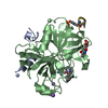
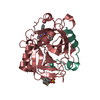
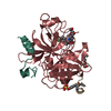
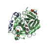
| Structure viewer | Molecule: MolmilJmol/JSmol |
|---|
- Downloads & links
-Download
| PDBx/mmCIF format | 3hat.cif.gz | 81 KB | Display | PDBx/mmCIF format |
|---|---|---|---|---|
| PDB format | pdb3hat.ent.gz | 58.6 KB | Display | PDB format |
| PDBx/mmJSON format | 3hat.json.gz | Tree view | PDBx/mmJSON format | |
| Others |  Other downloads Other downloads |
-Validation report
| Arichive directory | https://data.pdbj.org/pub/pdb/validation_reports/ha/3hatftp://data.pdbj.org/pub/pdb/validation_reports/ha/3hat | HTTPS FTP |
|---|
-Related structure data


-Links
 PDBj
PDBj


- Assembly
| Deposited unit | 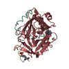
| ||||||||
|---|---|---|---|---|---|---|---|---|---|
| 1 |
| ||||||||
| Unit cell |
| ||||||||
| Atom site foot note | 1: CIS PROLINE - PRO H 37 | ||||||||
| Components on special symmetry positions |
|
-Components
| #1: Protein/peptide | Mass: 4096.534 Da / Num. of mol.: 1 / Source method: isolated from a natural source / Source: (natural) Homo sapiens (human) / Organ: BLOOD / Tissue: BLOOD / References: UniProt: P00734 |
|---|---|
| #2: Protein | Mass: 29780.219 Da / Num. of mol.: 1 / Source method: isolated from a natural source / Source: (natural) Homo sapiens (human) / Organ: BLOOD / Tissue: BLOOD / References: UniProt: P00734 |
| #3: Protein/peptide | Mass: 1534.554 Da / Num. of mol.: 1 / Source method: obtained synthetically / Source: (synth.) Hirudo medicinalis (medicinal leech) / References: UniProt: P09945 |
| #4: Protein/peptide | Mass: 537.632 Da / Num. of mol.: 1 / Source method: obtained synthetically Details: The capping residue of FPAM is not fully modeled. The SMILES string of the capping residue is OC(=O)C[C@H]1CCCN2N1C(=O)C(CCC2=O)C(NCC1=CC=CC=C1)C1=CC=CC=C1 Source: (synth.) synthetic construct (others) |
| #5: Water | ChemComp-HOH / Water Mass: 18.015 Da / Num. of mol.: 179 / Source method: isolated from a natural source / Formula: H2O Mass: 18.015 Da / Num. of mol.: 179 / Source method: isolated from a natural source / Formula: H2O |
| Compound details | THROMBIN IS CLEAVED BETWEEN RESIDUES 15 AND 16. CHAIN IDENTIFIER *L* IS USED FOR RESIDUES 1H - 15 ...THROMBIN IS CLEAVED BETWEEN RESIDUES 15 AND 16. CHAIN IDENTIFIER |
-Experimental details
-Experiment
| Experiment | Method: X-RAY DIFFRACTION |
|---|
- Sample preparation
| Crystal | Density Matthews: 2.56 Å3/Da / Density % sol: 51.89 % |
|---|---|
| Crystal grow | *PLUS Method: other / Details: Wu, T.P., (1993) Protein Eng., 6, 471. |
-Data collection
| Radiation | Scattering type: x-ray |
|---|---|
| Radiation wavelength | Relative weight: 1 |
| Reflection | *PLUS Highest resolution: 2.5 Å / Num. obs: 22675 / Num. measured all: 37533 / Rmerge(I) obs: 0.039 |
- Processing
| Software | Name: PROLSQ / Classification: refinement | |||||||||||||||||||||||||||||||||||||||||||||||||||||||||||||||
|---|---|---|---|---|---|---|---|---|---|---|---|---|---|---|---|---|---|---|---|---|---|---|---|---|---|---|---|---|---|---|---|---|---|---|---|---|---|---|---|---|---|---|---|---|---|---|---|---|---|---|---|---|---|---|---|---|---|---|---|---|---|---|---|---|
| Refinement | Rfactor obs: 0.14 / Highest resolution: 2.5 Å | |||||||||||||||||||||||||||||||||||||||||||||||||||||||||||||||
| Refinement step | Cycle: LAST / Highest resolution: 2.5 Å
| |||||||||||||||||||||||||||||||||||||||||||||||||||||||||||||||
| Refine LS restraints |
| |||||||||||||||||||||||||||||||||||||||||||||||||||||||||||||||
| Refinement | *PLUS Rfactor obs: 0.14 | |||||||||||||||||||||||||||||||||||||||||||||||||||||||||||||||
| Solvent computation | *PLUS | |||||||||||||||||||||||||||||||||||||||||||||||||||||||||||||||
| Displacement parameters | *PLUS | |||||||||||||||||||||||||||||||||||||||||||||||||||||||||||||||
| Refine LS restraints | *PLUS
|
