 Movie
Movie Controller

[English] 日本語
 Yorodumi
- PDB-3b7z: Crystal Structure of Yeast Sec14 Homolog Sfh1 in Complex with Pho... -
+ Open data

- Basic information
| Entry | Database: PDB / ID: 3b7z | ||||||
|---|---|---|---|---|---|---|---|
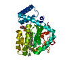
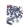
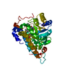
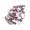
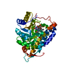
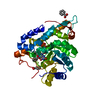
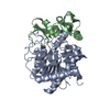
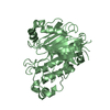
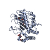
| Title | Crystal Structure of Yeast Sec14 Homolog Sfh1 in Complex with Phosphatidylcholine or Phosphatidylinositol | ||||||
 Components Components | Uncharacterized protein YKL091C | ||||||
 Keywords Keywords |  SIGNALING PROTEIN / Sec14 / Golgi / phospholipid / phosphatidylinositol / phosphatidycholine SIGNALING PROTEIN / Sec14 / Golgi / phospholipid / phosphatidylinositol / phosphatidycholine | ||||||
| Function / homology |  Function and homology information Function and homology information | ||||||
| Biological species |  Saccharomyces cerevisiae (brewer's yeast) Saccharomyces cerevisiae (brewer's yeast) | ||||||
| Method | X-RAY DIFFRACTION / SYNCHROTRON / MOLECULAR REPLACEMENT / molecular replacement / Resolution: 2.03 Å | ||||||
 Authors Authors | Ortlund, E.A. / Schaaf, G. / Redinbo, M.R. / Bankaitis, V. | ||||||
 Citation Citation | Journal: Mol.Cell / Year: 2008 Title: Functional anatomy of phospholipid binding and regulation of phosphoinositide homeostasis by proteins of the sec14 superfamily Authors: Schaaf, G. / Ortlund, E.A. / Tyeryar, K.R. / Mousley, C.J. / Ile, K.E. / Garrett, T.A. / Ren, J. / Woolls, M.J. / Raetz, C.R. / Redinbo, M.R. / Bankaitis, V.A. | ||||||
| History |
|
- Structure visualization
| Structure viewer | Molecule: MolmilJmol/JSmol |
|---|
- Downloads & links
-Download
| PDBx/mmCIF format | 3b7z.cif.gz | 86 KB | Display | PDBx/mmCIF format |
|---|---|---|---|---|
| PDB format | pdb3b7z.ent.gz | 62 KB | Display | PDB format |
| PDBx/mmJSON format | 3b7z.json.gz | Tree view | PDBx/mmJSON format | |
| Others |  Other downloads Other downloads |
-Validation report
| Arichive directory | https://data.pdbj.org/pub/pdb/validation_reports/b7/3b7zftp://data.pdbj.org/pub/pdb/validation_reports/b7/3b7z | HTTPS FTP |
|---|
-Related structure data


-Links
 PDBj
PDBj- Assembly
| Deposited unit | 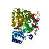
| ||||||||
|---|---|---|---|---|---|---|---|---|---|
| 1 |
| ||||||||
| Unit cell |
|
-Components
| #1: Protein | Mass: 37413.758 Da / Num. of mol.: 1 Source method: isolated from a genetically manipulated source Source: (gene. exp.) Saccharomyces cerevisiae (brewer's yeast)Strain: BY4741 / Plasmid: Pet28-SFH1 / Production host:  Escherichia coli (E. coli) / Strain (production host): BL21-CodonPlus (DE3)-RIL / References: UniProt: P33324 Escherichia coli (E. coli) / Strain (production host): BL21-CodonPlus (DE3)-RIL / References: UniProt: P33324 |
|---|---|
| #2: Chemical | ChemComp-B7N / (  Mass: 865.122 Da / Num. of mol.: 1 / Source method: obtained synthetically / Formula: C45H85O13P / Comment: phospholipid*YM Mass: 865.122 Da / Num. of mol.: 1 / Source method: obtained synthetically / Formula: C45H85O13P / Comment: phospholipid*YM |
| #3: Chemical | ChemComp-6PL / (  Mass: 763.100 Da / Num. of mol.: 1 / Source method: obtained synthetically / Formula: C42H85NO8P / Comment: phospholipid*YM Mass: 763.100 Da / Num. of mol.: 1 / Source method: obtained synthetically / Formula: C42H85NO8P / Comment: phospholipid*YM |
| #4: Water | ChemComp-HOH / Water Mass: 18.015 Da / Num. of mol.: 207 / Source method: isolated from a natural source / Formula: H2O Mass: 18.015 Da / Num. of mol.: 207 / Source method: isolated from a natural source / Formula: H2O |
-Experimental details
-Experiment
| Experiment | Method: X-RAY DIFFRACTION / Number of used crystals: 1 |
|---|
- Sample preparation
| Crystal | Density Matthews: 2.35 Å3/Da / Density % sol: 47.68 % |
|---|---|
| Crystal grow | Temperature: 295 K / Method: vapor diffusion, hanging drop / pH: 4.6 Details: 15-25% PEG 3350, 5% glycerol, and either 100 mM ammonium sulfate or 100mM potassium phosphate, 100 mM ammonium acetate, pH 4.6. Crystals were cryoprotected in 30% PEG 3350, 30% glycerol, and ...Details: 15-25% PEG 3350, 5% glycerol, and either 100 mM ammonium sulfate or 100mM potassium phosphate, 100 mM ammonium acetate, pH 4.6. Crystals were cryoprotected in 30% PEG 3350, 30% glycerol, and 200 mM ammonium sulfate, 100 mM ammonium acetate pH 4.6, VAPOR DIFFUSION, HANGING DROP, temperature 295K |
-Data collection
| Diffraction | Mean temperature: 100 K |
|---|---|
| Diffraction source | Source: SYNCHROTRON / Site: APS  / Beamline: 22-ID / Wavelength: 1 Å / Beamline: 22-ID / Wavelength: 1 Å |
| Detector | Type: MARMOSAIC 300 mm CCD / Detector: CCD / Date: Jun 14, 2006 |
| Radiation | Monochromator: Si 111 / Protocol: SINGLE WAVELENGTH / Monochromatic (M) / Laue (L): M / Scattering type: x-ray |
| Radiation wavelength | Wavelength: 1 Å / Relative weight: 1 |
| Reflection | Resolution: 2.03→50 Å / Num. obs: 22693 / % possible obs: 95.8 % / Observed criterion σ(F): 0 / Redundancy: 4.1 % / Rmerge(I) obs: 0.086 / Χ2: 1.315 / Net I/σ(I): 14.2 |
| Reflection shell | Resolution: 2.03→2.1 Å / Redundancy: 3.9 % / Rmerge(I) obs: 0.289 / Num. unique all: 2161 / Χ2: 1.468 / % possible all: 93.2 |
-Phasing
| Phasing | Method: molecular replacement |
|---|
- Processing
| Software |
| ||||||||||||||||||||||||||||||||||||
|---|---|---|---|---|---|---|---|---|---|---|---|---|---|---|---|---|---|---|---|---|---|---|---|---|---|---|---|---|---|---|---|---|---|---|---|---|---|
| Refinement | Method to determine structure: MOLECULAR REPLACEMENT / Resolution: 2.03→40.56 Å / Rfactor Rfree error: 0.006 / Data cutoff high absF: 146417.438 / Data cutoff low absF: 0 / Isotropic thermal model: RESTRAINED / Cross valid method: THROUGHOUT / σ(F): 0 / Stereochemistry target values: Engh & Huber
| ||||||||||||||||||||||||||||||||||||
| Solvent computation | Solvent model: FLAT MODEL / Bsol: 43.063 Å2 / ksol: 0.361 e/Å3 | ||||||||||||||||||||||||||||||||||||
| Displacement parameters | Biso mean: 31.4 Å2
| ||||||||||||||||||||||||||||||||||||
| Refine analyze |
| ||||||||||||||||||||||||||||||||||||
| Refinement step | Cycle: LAST / Resolution: 2.03→40.56 Å
| ||||||||||||||||||||||||||||||||||||
| Refine LS restraints |
| ||||||||||||||||||||||||||||||||||||
| LS refinement shell | Resolution: 2.03→2.16 Å / Rfactor Rfree error: 0.02 / Total num. of bins used: 6
| ||||||||||||||||||||||||||||||||||||
| Xplor file |
|
