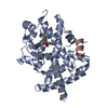
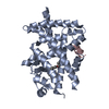
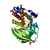
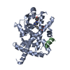
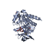
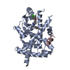
[English] 日本語
Yorodumi
- PDB-3b1m: Crystal structure of the PPARgamma-LBD complexed with a cercospor... -

+
Open data


ID or keywords:

Loading...

-
Basic information

Entry
Database: PDB / ID: 3b1m
TitleCrystal structure of the PPARgamma-LBD complexed with a cercosporamide derivative modulator Cerco-A
Components
  • Peroxisome proliferator-activated receptor gamma coactivator 1-alphaPeroxisome proliferator-activated receptor gamma
  • Peroxisome proliferator-activated receptor gamma
KeywordsTRANSCRIPTION
Function / homology
Function and homology information


positive regulation of mitochondrial DNA metabolic process / positive regulation of muscle tissue development / positive regulation of cellular respiration / positive regulation of fatty acid oxidation / : / : / lncRNA binding / cellular respiration / prostaglandin receptor activity / regulation of cholesterol transporter activity ...positive regulation of mitochondrial DNA metabolic process / positive regulation of muscle tissue development / positive regulation of cellular respiration / positive regulation of fatty acid oxidation / : / : / lncRNA binding / cellular respiration / prostaglandin receptor activity / regulation of cholesterol transporter activity / negative regulation of connective tissue replacement involved in inflammatory response wound healing / negative regulation of receptor signaling pathway via STAT / response to muscle activity / MECP2 regulates transcription factors / negative regulation of extracellular matrix assembly / negative regulation of vascular endothelial cell proliferation / negative regulation of cellular response to transforming growth factor beta stimulus / Activation of PPARGC1A (PGC-1alpha) by phosphorylation / negative regulation of cardiac muscle hypertrophy in response to stress / arachidonic acid binding / positive regulation of low-density lipoprotein receptor activity / positive regulation of adiponectin secretion / lipoprotein transport / negative regulation of sequestering of triglyceride / temperature homeostasis / positive regulation of vascular associated smooth muscle cell apoptotic process / macrophage derived foam cell differentiation / DNA binding domain binding / STAT family protein binding / positive regulation of fatty acid metabolic process / response to lipid / negative regulation of SMAD protein signal transduction / positive regulation of ATP biosynthetic process / response to starvation / LBD domain binding / negative regulation of type II interferon-mediated signaling pathway / negative regulation of cholesterol storage / response to dietary excess / E-box binding / alpha-actinin binding / negative regulation of blood vessel endothelial cell migration / lipid homeostasis / intracellular glucose homeostasis / fatty acid oxidation / negative regulation of vascular associated smooth muscle cell proliferation / R-SMAD binding / monocyte differentiation / positive regulation of cholesterol efflux / cellular response to low-density lipoprotein particle stimulus / negative regulation of macrophage derived foam cell differentiation / negative regulation of lipid storage / negative regulation of BMP signaling pathway / white fat cell differentiation / negative regulation of mitochondrial fission / positive regulation of fat cell differentiation / negative regulation of osteoblast differentiation / long-chain fatty acid transport / retinoic acid receptor signaling pathway / positive regulation of DNA binding / cell fate commitment / BMP signaling pathway / adipose tissue development / FOXO-mediated transcription of oxidative stress, metabolic and neuronal genes / nuclear retinoid X receptor binding / energy homeostasis / brown fat cell differentiation / negative regulation of signaling receptor activity / regulation of cellular response to insulin stimulus / cell maturation / positive regulation of gluconeogenesis / digestion / positive regulation of adipose tissue development / epithelial cell differentiation / peroxisome proliferator activated receptor signaling pathway / mitochondrion organization / respiratory electron transport chain / hormone-mediated signaling pathway / negative regulation of angiogenesis / response to nutrient / negative regulation of MAP kinase activity / RNA splicing / fatty acid metabolic process / nuclear receptor coactivator activity / SUMOylation of transcription cofactors / negative regulation of miRNA transcription / placenta development / Regulation of PTEN gene transcription / negative regulation of smooth muscle cell proliferation / gluconeogenesis / transcription coregulator binding / transcription initiation at RNA polymerase II promoter / nuclear receptor binding / transcription coregulator activity / peptide binding / negative regulation of transforming growth factor beta receptor signaling pathway / circadian regulation of gene expression / Heme signaling / SUMOylation of intracellular receptors / mRNA transcription by RNA polymerase II / Transcriptional activation of mitochondrial biogenesis
Similarity search - Function
PGC-1alpha, RNA recognition motif / PGC-1 / Peroxisome proliferator-activated receptor gamma / Peroxisome proliferator-activated receptor gamma, N-terminal / PPAR gamma N-terminal region / Peroxisome proliferator-activated receptor / Retinoid X Receptor / Retinoid X Receptor / RNA recognition motif / RNA recognition motif ...PGC-1alpha, RNA recognition motif / PGC-1 / Peroxisome proliferator-activated receptor gamma / Peroxisome proliferator-activated receptor gamma, N-terminal / PPAR gamma N-terminal region / Peroxisome proliferator-activated receptor / Retinoid X Receptor / Retinoid X Receptor / RNA recognition motif / RNA recognition motif / Eukaryotic RNA Recognition Motif (RRM) profile. / RNA recognition motif domain / RNA-binding domain superfamily / Nuclear hormone receptor / Nuclear hormones receptors DNA-binding region signature. / Zinc finger, nuclear hormone receptor-type / Zinc finger, C4 type (two domains) / Nuclear hormone receptors DNA-binding domain profile. / c4 zinc finger in nuclear hormone receptors / Nuclear hormone receptor, ligand-binding domain / Nuclear hormone receptor-like domain superfamily / Ligand-binding domain of nuclear hormone receptor / Nuclear receptor (NR) ligand-binding (LBD) domain profile. / Ligand binding domain of hormone receptors / Zinc finger, NHR/GATA-type / Nucleotide-binding alpha-beta plait domain superfamily / Orthogonal Bundle / Mainly Alpha
Similarity search - Domain/homology
Chem-KRC / Peroxisome proliferator-activated receptor gamma / Peroxisome proliferator-activated receptor gamma coactivator 1-alpha
Similarity search - Component
Biological speciesHomo sapiens (human)
MethodX-RAY DIFFRACTION / MOLECULAR REPLACEMENT / Resolution: 1.6 Å
AuthorsMatsui, Y. / Hiroyuki, H.
CitationJournal: Biol.Pharm.Bull. / Year: 2011
Title: Pharmacology and in Vitro Profiling of a Novel Peroxisome Proliferator-Activated Receptor gamma Ligand, Cerco-A
Authors: Wakabayashi, K. / Hayashi, S. / Matsui, Y. / Matsumoto, T. / Furukawa, A. / Kuroha, M. / Tanaka, N. / Inaba, T. / Kanda, S. / Tanaka, J. / Okuyama, R. / Wakimoto, S. / Ogata, T. / Araki, K. / Ohsumi, J.
History
DepositionJul 5, 2011Deposition site: PDBJ / Processing site: PDBJ
Revision 1.0Aug 24, 2011Provider: repository / Type: Initial release
Revision 1.1Nov 1, 2023Group: Data collection / Database references ...Data collection / Database references / Derived calculations / Refinement description
Category: chem_comp_atom / chem_comp_bond ...chem_comp_atom / chem_comp_bond / database_2 / pdbx_initial_refinement_model / struct_ref_seq_dif / struct_site
Item: _database_2.pdbx_DOI / _database_2.pdbx_database_accession ..._database_2.pdbx_DOI / _database_2.pdbx_database_accession / _struct_ref_seq_dif.details / _struct_site.pdbx_auth_asym_id / _struct_site.pdbx_auth_comp_id / _struct_site.pdbx_auth_seq_id

-
Structure visualization

Structure viewerMolecule:
MolmilJmol/JSmol

Downloads & links

-
Assembly

Deposited unit
A: Peroxisome proliferator-activated receptor gamma
B: Peroxisome proliferator-activated receptor gamma coactivator 1-alpha
hetero molecules


Theoretical massNumber of molelcules
Total (without water)34,9933
Polymers34,4802
Non-polymers5141
Water5,008278
1


  • Idetical with deposited unit
  • defined by author&software
TypeNameSymmetry operationNumber
identity operation1_555x,y,z1
Buried area1020 Å2
ΔGint-9 kcal/mol
Surface area13370 Å2
MethodPISA
Unit cell
Length a, b, c (Å)38.946, 54.622, 66.782
Angle α, β, γ (deg.)90.00, 92.21, 90.00
Int Tables number4
Space group name H-MP1211

-
Components

#1: Protein Peroxisome proliferator-activated receptor gamma / / PPAR-gamma / Nuclear receptor subfamily 1 group C member 3


Mass: 32412.576 Da / Num. of mol.: 1 / Fragment: ligand binding domain, UNP residues 234-505
Source method: isolated from a genetically manipulated source
Source: (gene. exp.) Homo sapiens (human) / Gene: PPARG, NR1C3 / Plasmid: pQE-30 / Production host: Escherichia coli (E. coli) / References: UniProt: P37231
#2: Protein/peptide Peroxisome proliferator-activated receptor gamma coactivator 1-alpha / Peroxisome proliferator-activated receptor gamma / PGC-1-alpha / PPAR-gamma coactivator 1-alpha / PPARGC-1-alpha / Ligand effect modulator 6


Mass: 2067.381 Da / Num. of mol.: 1
Fragment: peptide containing LXXLL box, UNP residues 136-154
Source method: obtained synthetically / Details: This sequence occurs naturally in humans. / Source: (synth.) Homo sapiens (human) / References: UniProt: Q9UBK2
#3: Chemical ChemComp-KRC / (9aS)-8-acetyl-N-[(2-ethylnaphthalen-1-yl)methyl]-1,7-dihydroxy-3-methoxy-9a-methyl-9-oxo-9,9a-dihydrodibenzo[b,d]furan-4-carboxamide


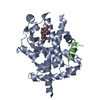
Mass: 513.538 Da / Num. of mol.: 1 / Source method: obtained synthetically / Formula: C30H27NO7
#4: Water ChemComp-HOH / water / Water


Mass: 18.015 Da / Num. of mol.: 278 / Source method: isolated from a natural source / Formula: H2O

-
Experimental details

-
Experiment

ExperimentMethod: X-RAY DIFFRACTION / Number of used crystals: 1

-
Sample preparation

CrystalDensity Matthews: 2.06 Å3/Da / Density % sol: 40.25 %
Crystal growTemperature: 295 K / Method: vapor diffusion, hanging drop / pH: 8.5
Details: PEG 4000, sodium thiocyanate, Tris-hydrochloride, pH 8.5, VAPOR DIFFUSION, HANGING DROP, temperature 295K

-
Data collection

DiffractionMean temperature: 93 K
Diffraction sourceSource: ROTATING ANODE / Type: RIGAKU FR-E DW / Wavelength: 1.5418 Å
DetectorType: RIGAKU RAXIS VII / Detector: IMAGE PLATE / Date: Jun 28, 2005
RadiationProtocol: SINGLE WAVELENGTH / Monochromatic (M) / Laue (L): M / Scattering type: x-ray
Radiation wavelengthWavelength: 1.5418 Å / Relative weight: 1
ReflectionResolution: 1.6→50 Å / Num. all: 37106 / Num. obs: 36868 / % possible obs: 99.4 % / Biso Wilson estimate: 22.8 Å2 / Rsym value: 0.055
Reflection shellResolution: 1.6→1.66 Å / Mean I/σ(I) obs: 4.25 / Num. unique all: 3530 / Rsym value: 0.255 / % possible all: 96.2

-
Processing

Software
NameClassification
CrystalCleardata collection
CNSrefinement
HKL-2000data reduction
HKL-2000data scaling
CNSphasing
RefinementMethod to determine structure: MOLECULAR REPLACEMENT
Starting model: PDB ENTRY 3LMP
Resolution: 1.6→19 Å / Rfactor Rfree error: 0.004 / Data cutoff high absF: 867084.52 / Data cutoff low absF: 0 / Isotropic thermal model: RESTRAINED / Cross valid method: THROUGHOUT / σ(F): 0 / Stereochemistry target values: Engh & Huber / Details: BULK SOLVENT MODEL USED
RfactorNum. reflection% reflectionSelection details
Rfree0.244 3682 10 %RANDOM
Rwork0.217 ---
obs0.217 36852 99.3 %-
Solvent computationSolvent model: FLAT MODEL / Bsol: 49.0583 Å2 / ksol: 0.4 e/Å3
Displacement parametersBiso mean: 22.8 Å2
Baniso -1Baniso -2Baniso -3
1-3.72 Å20 Å2-1.57 Å2
2---1.26 Å20 Å2
3----2.46 Å2
Refine analyze
FreeObs
Luzzati coordinate error0.24 Å0.2 Å
Luzzati d res low-5 Å
Luzzati sigma a0.08 Å0.07 Å
Refinement stepCycle: LAST / Resolution: 1.6→19 Å
ProteinNucleic acidLigandSolventTotal
Num. atoms2166 0 38 278 2482
Refine LS restraints
Refine-IDTypeDev idealDev ideal target
X-RAY DIFFRACTIONc_bond_d0.005
X-RAY DIFFRACTIONc_bond_d_na
X-RAY DIFFRACTIONc_bond_d_prot
X-RAY DIFFRACTIONc_angle_d
X-RAY DIFFRACTIONc_angle_d_na
X-RAY DIFFRACTIONc_angle_d_prot
X-RAY DIFFRACTIONc_angle_deg1
X-RAY DIFFRACTIONc_angle_deg_na
X-RAY DIFFRACTIONc_angle_deg_prot
X-RAY DIFFRACTIONc_dihedral_angle_d18.3
X-RAY DIFFRACTIONc_dihedral_angle_d_na
X-RAY DIFFRACTIONc_dihedral_angle_d_prot
X-RAY DIFFRACTIONc_improper_angle_d0.72
X-RAY DIFFRACTIONc_improper_angle_d_na
X-RAY DIFFRACTIONc_improper_angle_d_prot
X-RAY DIFFRACTIONc_mcbond_it1.171.5
X-RAY DIFFRACTIONc_mcangle_it1.882
X-RAY DIFFRACTIONc_scbond_it1.972
X-RAY DIFFRACTIONc_scangle_it2.982.5
LS refinement shellResolution: 1.6→1.7 Å / Rfactor Rfree error: 0.015 / Total num. of bins used: 6
RfactorNum. reflection% reflection
Rfree0.352 579 9.7 %
Rwork0.331 5402 -
obs-5402 97.3 %
Xplor file
Refine-IDSerial noParam fileTopol file
X-RAY DIFFRACTION1protein_rep.paramprotein.top
X-RAY DIFFRACTION2water.paramwater.top
X-RAY DIFFRACTION3CRC.paramCRC.top

+
About Yorodumi

-
News

-
Feb 9, 2022. New format data for meta-information of EMDB entries

New format data for meta-information of EMDB entries

  • Version 3 of the EMDB header file is now the official format.
  • The previous official version 1.9 will be removed from the archive.

Related info.:EMDB header

External links:wwPDB to switch to version 3 of the EMDB data model

-
Aug 12, 2020. Covid-19 info

Covid-19 info

URL: https://pdbj.org/emnavi/covid19.php

New page: Covid-19 featured information page in EM Navigator.

Related info.:Covid-19 info / Mar 5, 2020. Novel coronavirus structure data

+
Mar 5, 2020. Novel coronavirus structure data

Novel coronavirus structure data

Related info.:Yorodumi Speices / Aug 12, 2020. Covid-19 info

External links:COVID-19 featured content - PDBj / Molecule of the Month (242):Coronavirus Proteases

+
Jan 31, 2019. EMDB accession codes are about to change! (news from PDBe EMDB page)

EMDB accession codes are about to change! (news from PDBe EMDB page)

  • The allocation of 4 digits for EMDB accession codes will soon come to an end. Whilst these codes will remain in use, new EMDB accession codes will include an additional digit and will expand incrementally as the available range of codes is exhausted. The current 4-digit format prefixed with “EMD-” (i.e. EMD-XXXX) will advance to a 5-digit format (i.e. EMD-XXXXX), and so on. It is currently estimated that the 4-digit codes will be depleted around Spring 2019, at which point the 5-digit format will come into force.
  • The EM Navigator/Yorodumi systems omit the EMD- prefix.

Related info.:Q: What is EMD? / ID/Accession-code notation in Yorodumi/EM Navigator

External links:EMDB Accession Codes are Changing Soon! / Contact to PDBj

+
Jul 12, 2017. Major update of PDB

Major update of PDB

  • wwPDB released updated PDB data conforming to the new PDBx/mmCIF dictionary.
  • This is a major update changing the version number from 4 to 5, and with Remediation, in which all the entries are updated.
  • In this update, many items about electron microscopy experimental information are reorganized (e.g. em_software).
  • Now, EM Navigator and Yorodumi are based on the updated data.

External links:wwPDB Remediation / Enriched Model Files Conforming to OneDep Data Standards Now Available in the PDB FTP Archive

-
Yorodumi

Thousand views of thousand structures

  • Yorodumi is a browser for structure data from EMDB, PDB, SASBDB, etc.
  • This page is also the successor to EM Navigator detail page, and also detail information page/front-end page for Omokage search.
  • The word "yorodu" (or yorozu) is an old Japanese word meaning "ten thousand". "mi" (miru) is to see.

Related info.:EMDB / PDB / SASBDB / Comparison of 3 databanks / Yorodumi Search / Aug 31, 2016. New EM Navigator & Yorodumi / Yorodumi Papers / Jmol/JSmol / Function and homology information / Changes in new EM Navigator and Yorodumi

Read more