| Entry | Database: PDB / ID: 6k0t
|
|---|
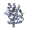
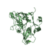
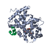
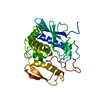
| Title | Crystal Structure of PPARgamma Ligand Binding Domain in complex with dibenzooxepine derivative compound-17 |
|---|
 Components Components | - Peroxisome proliferator-activated receptor gamma
- Peroxisome proliferator-activated receptor gamma coactivator 1-alpha
|
|---|
 Keywords Keywords | NUCLEAR PROTEIN / PPARgamma / Ligand binding domain / PPARg modulator / Cancer |
|---|
| Function / homology |  Function and homology information Function and homology information
Regulation of MITF-M dependent genes involved in metabolism / positive regulation of fatty acid oxidation / Activation of PPARGC1A (PGC-1alpha) by phosphorylation / prostaglandin receptor activity / negative regulation of receptor signaling pathway via STAT / cellular respiration / MECP2 regulates transcription factors / beige fat cell differentiation / negative regulation of vascular endothelial cell proliferation / negative regulation of extracellular matrix assembly ...Regulation of MITF-M dependent genes involved in metabolism / positive regulation of fatty acid oxidation / Activation of PPARGC1A (PGC-1alpha) by phosphorylation / prostaglandin receptor activity / negative regulation of receptor signaling pathway via STAT / cellular respiration / MECP2 regulates transcription factors / beige fat cell differentiation / negative regulation of vascular endothelial cell proliferation / negative regulation of extracellular matrix assembly / negative regulation of connective tissue replacement involved in inflammatory response wound healing / positive regulation of cholesterol transport / negative regulation of cellular response to transforming growth factor beta stimulus / arachidonate binding / positive regulation of adiponectin secretion / DNA binding domain binding / positive regulation of vascular associated smooth muscle cell apoptotic process / negative regulation of cardiac muscle hypertrophy in response to stress / positive regulation of fatty acid metabolic process / positive regulation of lipid metabolic process / STAT family protein binding / response to muscle activity / response to starvation / WW domain binding / negative regulation of type II interferon-mediated signaling pathway / LBD domain binding / negative regulation of cholesterol storage / response to lipid / positive regulation of lipoprotein transport / response to dietary excess / lncRNA binding / negative regulation of SMAD protein signal transduction / fatty acid oxidation / lipid homeostasis / E-box binding / temperature homeostasis / adipose tissue development / R-SMAD binding / negative regulation of blood vessel endothelial cell migration / white fat cell differentiation / monocyte differentiation / negative regulation of vascular associated smooth muscle cell proliferation / negative regulation of BMP signaling pathway / alpha-actinin binding / negative regulation of macrophage derived foam cell differentiation / negative regulation of lipid storage / BMP signaling pathway / positive regulation of cholesterol efflux / cell fate commitment / cellular response to low-density lipoprotein particle stimulus / FOXO-mediated transcription of oxidative stress, metabolic and neuronal genes / long-chain fatty acid transport / negative regulation of mitochondrial fission / fat cell differentiation / nuclear retinoid X receptor binding / negative regulation of osteoblast differentiation / positive regulation of fat cell differentiation / retinoic acid receptor signaling pathway / energy homeostasis / Transcriptional regulation of brown and beige adipocyte differentiation by EBF2 / digestion / intracellular receptor signaling pathway / peroxisome proliferator activated receptor signaling pathway / hormone-mediated signaling pathway / cell maturation / negative regulation of MAPK cascade / positive regulation of adipose tissue development / peptide binding / epithelial cell differentiation / regulation of cellular response to insulin stimulus / response to nutrient / RORA,B,C and NR1D1 (REV-ERBA) regulate gene expression / SUMOylation of transcription cofactors / Expression of BMAL (ARNTL), CLOCK, and NPAS2 / negative regulation of miRNA transcription / placenta development / negative regulation of angiogenesis / positive regulation of gluconeogenesis / brown fat cell differentiation / fatty acid metabolic process / intracellular glucose homeostasis / RNA splicing / positive regulation of apoptotic signaling pathway / nuclear receptor binding / Regulation of PTEN gene transcription / transcription coregulator binding / gluconeogenesis / SUMOylation of intracellular receptors / respiratory electron transport chain / circadian regulation of gene expression / transcription coregulator activity / negative regulation of smooth muscle cell proliferation / mitochondrion organization / Heme signaling / negative regulation of transforming growth factor beta receptor signaling pathway / transcription initiation at RNA polymerase II promoter / PPARA activates gene expression / Transcriptional activation of mitochondrial biogenesis / Nuclear Receptor transcription pathway / Transcriptional regulation of white adipocyte differentiationSimilarity search - Function PGC-1alpha, RNA recognition motif / PGC-1 / Peroxisome proliferator-activated receptor gamma / Peroxisome proliferator-activated receptor gamma, N-terminal / PPAR gamma N-terminal region / Peroxisome proliferator-activated receptor / : / Retinoid X Receptor / Retinoid X Receptor / RNA recognition motif ...PGC-1alpha, RNA recognition motif / PGC-1 / Peroxisome proliferator-activated receptor gamma / Peroxisome proliferator-activated receptor gamma, N-terminal / PPAR gamma N-terminal region / Peroxisome proliferator-activated receptor / : / Retinoid X Receptor / Retinoid X Receptor / RNA recognition motif / RNA recognition motif / Eukaryotic RNA Recognition Motif (RRM) profile. / RNA recognition motif domain / RNA-binding domain superfamily / Nuclear hormone receptor / Nuclear hormones receptors DNA-binding region signature. / Zinc finger, nuclear hormone receptor-type / Double treble clef zinc finger, C4 type / Nuclear hormone receptors DNA-binding domain profile. / c4 zinc finger in nuclear hormone receptors / Nuclear hormone receptor, ligand-binding domain / Nuclear hormone receptor-like domain superfamily / Ligand-binding domain of nuclear hormone receptor / Nuclear receptor (NR) ligand-binding (LBD) domain profile. / Ligand binding domain of hormone receptors / Zinc finger, NHR/GATA-type / Nucleotide-binding alpha-beta plait domain superfamily / Orthogonal Bundle / Mainly AlphaSimilarity search - Domain/homology Chem-CTU / Peroxisome proliferator-activated receptor gamma / Peroxisome proliferator-activated receptor gamma coactivator 1-alphaSimilarity search - Component |
|---|
| Biological species |  Homo sapiens (human) Homo sapiens (human) |
|---|
| Method |  X-RAY DIFFRACTION / SYNCHROTRON / MOLECULAR REPLACEMENT / Resolution: 1.84 Å X-RAY DIFFRACTION / SYNCHROTRON / MOLECULAR REPLACEMENT / Resolution: 1.84 Å |
|---|
 Authors Authors | Suzuki, M. / Yamamoto, K. / Takahashi, Y. / Saito, J. |
|---|
 Citation Citation | Journal: Bioorg.Med.Chem. / Year: 2019
Title: Development of a novel class of peroxisome proliferator-activated receptor (PPAR) gamma ligands as an anticancer agent with a unique binding mode based on a non-thiazolidinedione scaffold.
Authors: Yamamoto, K. / Tamura, T. / Nakamura, R. / Hosoe, S. / Matsubara, M. / Nagata, K. / Kodaira, H. / Uemori, T. / Takahashi, Y. / Suzuki, M. / Saito, J.I. / Ueno, K. / Shuto, S. |
|---|
| History | | Deposition | May 7, 2019 | Deposition site: PDBJ / Processing site: PDBJ |
|---|
| Revision 1.0 | Oct 30, 2019 | Provider: repository / Type: Initial release |
|---|
| Revision 1.1 | Nov 6, 2019 | Group: Data collection / Database references / Category: citation / Item: _citation.journal_volume |
|---|
| Revision 1.2 | Nov 6, 2024 | Group: Data collection / Database references / Structure summary
Category: chem_comp_atom / chem_comp_bond ...chem_comp_atom / chem_comp_bond / database_2 / pdbx_entry_details / pdbx_modification_feature
Item: _database_2.pdbx_DOI / _database_2.pdbx_database_accession |
|---|
|
|---|
 Movie
Movie Controller

 Yorodumi
 Open data
 Basic information
 Structure visualization
 Downloads & links
 Other downloads
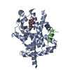


 PDBj
PDBj


 Assembly


 Mass: 528.961 Da / Num. of mol.: 2 / Source method: obtained synthetically / Formula: C29H22ClFN4O3 / Feature type: SUBJECT OF INVESTIGATION
 Mass: 18.015 Da / Num. of mol.: 449 / Source method: isolated from a natural source / Formula: H2O
 Sample preparation
 / Beamline: BL-17A / Wavelength: 0.98 Å
 Processing